


Crystal structure of Ferredoxin (flavodoxin):NADP(+) oxidoreductase from Klebsiella pneumoniae
Lovell, S., Liu, L., Seibold, S., Battaile, K.P.To be published.
Experimental Data Snapshot
Entity ID: 1 | |||||
|---|---|---|---|---|---|
| Molecule | Chains | Sequence Length | Organism | Details | Image |
| Flavodoxin/ferredoxin--NADP reductase | 256 | Klebsiella pneumoniae | Mutation(s): 0 Gene Names: fpr, BL124_00025130, DRB11_17480, EAO17_13200, G7Z27_00550, GJJ08_000550, GNE24_19530, GNG14_18110, SAMEA3499874_03593, SAMEA3500057_03748... EC: 1.18.1.2 (PDB Primary Data), 1.19.1.1 (PDB Primary Data) | 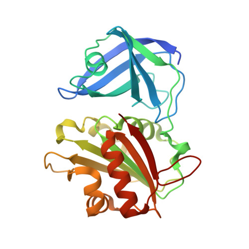 | |
UniProt | |||||
Entity Groups | |||||
| Sequence Clusters | 30% Identity50% Identity70% Identity90% Identity95% Identity100% Identity | ||||
| UniProt Group | A0A0G3NED2 | ||||
Sequence AnnotationsExpand | |||||
Reference Sequence | |||||
| Ligands 5 Unique | |||||
|---|---|---|---|---|---|
| ID | Chains | Name / Formula / InChI Key | 2D Diagram | 3D Interactions | |
| FAD (Subject of Investigation/LOI) Download:Ideal Coordinates CCD File | C [auth A], H [auth B] | FLAVIN-ADENINE DINUCLEOTIDE C27 H33 N9 O15 P2 VWWQXMAJTJZDQX-UYBVJOGSSA-N |  | ||
| NAP (Subject of Investigation/LOI) Download:Ideal Coordinates CCD File | D [auth A], I [auth B] | NADP NICOTINAMIDE-ADENINE-DINUCLEOTIDE PHOSPHATE C21 H28 N7 O17 P3 XJLXINKUBYWONI-NNYOXOHSSA-N |  | ||
| 1PE Download:Ideal Coordinates CCD File | E [auth A], J [auth B] | PENTAETHYLENE GLYCOL C10 H22 O6 JLFNLZLINWHATN-UHFFFAOYSA-N |  | ||
| CA Download:Ideal Coordinates CCD File | F [auth A], K [auth B] | CALCIUM ION Ca BHPQYMZQTOCNFJ-UHFFFAOYSA-N |  | ||
| MG Download:Ideal Coordinates CCD File | G [auth A] | MAGNESIUM ION Mg JLVVSXFLKOJNIY-UHFFFAOYSA-N |  | ||
| Length ( Å ) | Angle ( ˚ ) |
|---|---|
| a = 47.24 | α = 106.69 |
| b = 56.02 | β = 97.49 |
| c = 56.11 | γ = 83.4 |
| Software Name | Purpose |
|---|---|
| XDS | data reduction |
| Aimless | data scaling |
| PHASER | phasing |
| PHENIX | refinement |
| PDB_EXTRACT | data extraction |
| Funding Organization | Location | Grant Number |
|---|---|---|
| National Institutes of Health/National Institute Of Allergy and Infectious Diseases (NIH/NIAID) | United States | HHSN272201700059C |
| National Institutes of Health/Office of the Director | United States | S10OD030394 |


