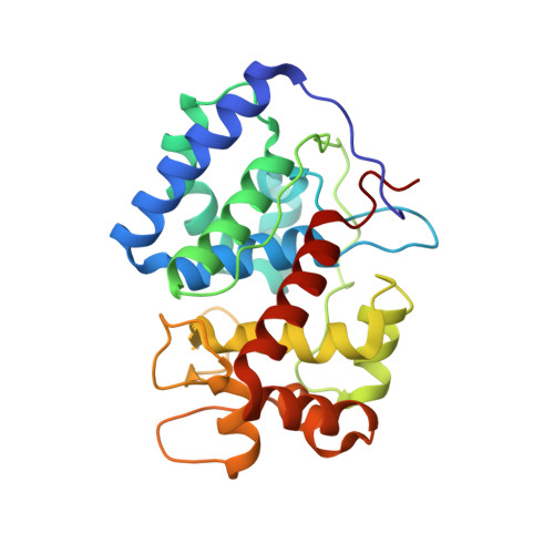
8DJT

Cytosolic ascorbate peroxidase from Sorghum bicolor - four ascorbates complex


Experimental Data Snapshot

  • Method: X-RAY DIFFRACTION
  • Resolution: 1.16 Å
  • R-Value Free: 0.151 
  • R-Value Work: 0.138 
  • R-Value Observed: 0.138 

Starting Model: experimental
View more details

wwPDB Validation   3D Report Full Report


Ligand Structure Quality Assessment 


This is version 1.2 of the entry. See complete history


Literature

A sorghum ascorbate peroxidase with four binding sites has activity against ascorbate and phenylpropanoids.

Zhang, B.Lewis, J.A.Vermerris, W.Sattler, S.E.Kang, C.

(2023) Plant Physiol 192: 102-118

  • DOI: https://doi.org/10.1093/plphys/kiac604
  • Primary Citation of Related Structures:  
    8DJR, 8DJS, 8DJT, 8DJU, 8DJW, 8DJX

  • PubMed Abstract: 

    In planta, H2O2 is produced as a by-product of enzymatic reactions and during defense responses. Ascorbate peroxidase (APX) is a key enzyme involved in scavenging cytotoxic H2O2. Here, we report the crystal structure of cytosolic APX from sorghum (Sorghum bicolor) (Sobic.001G410200). While the overall structure of SbAPX was similar to that of other APXs, SbAPX uniquely displayed four bound ascorbates rather than one. In addition to the ɣ-heme pocket identified in other APXs, ascorbates were bound at the δ-meso and two solvent-exposed pockets. Consistent with the presence of multiple binding sites, our results indicated that the H2O2-dependent oxidation of ascorbate displayed positive cooperativity. Bound ascorbate at two surface sites established an intricate proton network with ascorbate at the ɣ-heme edge and δ-meso sites. Based on crystal structures, steady-state kinetics, and site-directed mutagenesis results, both ascorbate molecules at the ɣ-heme edge and the one at the surface are expected to participate in the oxidation reaction. We provide evidence that the H2O2-dependent oxidation of ascorbate by APX produces a C2-hydrated bicyclic hemiketal form of dehydroascorbic acid at the ɣ-heme edge, indicating two successive electron transfers from a single-bound ascorbate. In addition, the δ-meso site was shared with several organic compounds, including p-coumaric acid and other phenylpropanoids, for the potential radicalization reaction. Site-directed mutagenesis of the critical residue at the ɣ-heme edge (R172A) only partially reduced polymerization activity. Thus, APX removes stress-generated H2O2 with ascorbates, and also uses this same H2O2 to potentially fortify cell walls via oxidative polymerization of phenylpropanoids in response to stress.


  • Organizational Affiliation

    Department of Chemistry, Washington State University, Pullman, Washington 99164, USA.


Macromolecules
Find similar proteins by:  (by identity cutoff)  |  3D Structure
Entity ID: 1
MoleculeChains Sequence LengthOrganismDetailsImage
L-ascorbate peroxidase250Sorghum bicolorMutation(s): 0 
Gene Names: SORBI_3001G410200
EC: 1.11.1.11
UniProt
Find proteins for C5WNL8 (Sorghum bicolor)
Explore C5WNL8 
Go to UniProtKB:  C5WNL8
Entity Groups  
Sequence Clusters30% Identity50% Identity70% Identity90% Identity95% Identity100% Identity
UniProt GroupC5WNL8
Sequence Annotations
Expand
  • Reference Sequence
Small Molecules
Ligands 4 Unique
IDChains Name / Formula / InChI Key2D Diagram3D Interactions
HEM
Query on HEM

Download Ideal Coordinates CCD File 
B [auth A]PROTOPORPHYRIN IX CONTAINING FE
C34 H32 Fe N4 O4
KABFMIBPWCXCRK-RGGAHWMASA-L
ASC (Subject of Investigation/LOI)
Query on ASC

Download Ideal Coordinates CCD File 
C [auth A],
D [auth A],
F [auth A],
G [auth A]
ASCORBIC ACID
C6 H8 O6
CIWBSHSKHKDKBQ-JLAZNSOCSA-N
GOL
Query on GOL

Download Ideal Coordinates CCD File 
E [auth A]GLYCEROL
C3 H8 O3
PEDCQBHIVMGVHV-UHFFFAOYSA-N
NA
Query on NA

Download Ideal Coordinates CCD File 
H [auth A]SODIUM ION
Na
FKNQFGJONOIPTF-UHFFFAOYSA-N
Experimental Data & Validation

Experimental Data

  • Method: X-RAY DIFFRACTION
  • Resolution: 1.16 Å
  • R-Value Free: 0.151 
  • R-Value Work: 0.138 
  • R-Value Observed: 0.138 
  • Space Group: P 21 21 21
Unit Cell:
Length ( Å )Angle ( ˚ )
a = 38.296α = 90
b = 75.097β = 90
c = 82.997γ = 90
Software Package:
Software NamePurpose
PHENIXrefinement
HKL-2000data scaling
PDB_EXTRACTdata extraction
XDSdata reduction
PHENIXphasing

Structure Validation

View Full Validation Report


Ligand Structure Quality Assessment 


Entry History & Funding Information

Deposition Data


Funding OrganizationLocationGrant Number
National Science Foundation (NSF, United States)United StatesCHE 1804699
National Science Foundation (NSF, United States)United StatesMCB-2043248

Revision History  (Full details and data files)

  • Version 1.0: 2023-01-18
    Type: Initial release
  • Version 1.1: 2023-05-17
    Changes: Database references
  • Version 1.2: 2023-10-25
    Changes: Data collection, Refinement description