N -Heterocyclic 3-Pyridyl Carboxamide Inhibitors of DHODH for the Treatment of Acute Myelogenous Leukemia.
Cisar, J.S., Pietsch, C., DeRatt, L.G., Jacoby, E., Kazmi, F., Keohane, C., Legenski, K., Matico, R., Shaffer, P., Simonnet, Y., Tanner, A., Wang, C.Y., Wang, W., Attar, R., Edwards, J.P., Kuduk, S.D.(2022) J Med Chem 65: 11241-11256
- PubMed: 35925768 Search on PubMed
- DOI: https://doi.org/10.1021/acs.jmedchem.2c00788
- Primary Citation Related Structures:
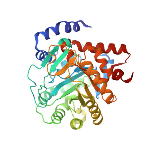
8DHF, 8DHG, 8DHH - PubMed Abstract:
Acute myelogenous leukemia (AML), a disease of the blood and bone marrow, is characterized by the inability of myeloblasts to differentiate into mature cell types. Dihydroorotate dehydrogenase (DHODH) is an enzyme well-known in the pyrimidine biosynthesis pathway; however, small molecule DHODH inhibitors were recently shown to induce differentiation in multiple AML subtypes. Using virtual screening and structure-based drug design approaches, a new series of N-heterocyclic 3-pyridyl carboxamide DHODH inhibitors were discovered. Two lead compounds, 19 and 29 , have potent biochemical and cellular DHODH activity, favorable physicochemical properties, and efficacy in a preclinical model of AML.
- Janssen Research and Development, 1400 McKean Rd, Spring House, Pennsylvania 19477, United States.
Organizational Affiliation: