8DGG

Structure of glycosylated LAG-3 homodimer


Experimental Data Snapshot

  • Method: X-RAY DIFFRACTION
  • Resolution: 3.78 Å
  • R-Value Free: 0.303 
  • R-Value Work: 0.264 
  • R-Value Observed: 0.266 

Starting Model: in silico
View more details

wwPDB Validation   3D Report Full Report


Ligand Structure Quality Assessment 


This is version 2.0 of the entry. See complete history


Literature

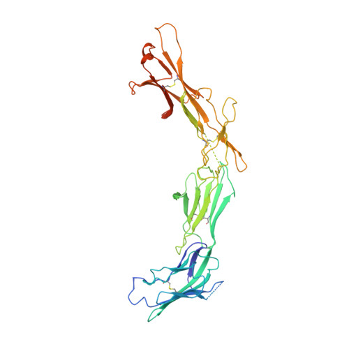
Structural insights reveal interplay between LAG-3 homodimerization, ligand binding, and function.

Silberstein, J.L.Du, J.Chan, K.W.Frank, J.A.Mathews, I.I.Kim, Y.B.You, J.Lu, Q.Liu, J.Philips, E.A.Liu, P.Rao, E.Fernandez, D.Rodriguez, G.E.Kong, X.P.Wang, J.Cochran, J.R.

(2024) Proc Natl Acad Sci U S A 121: e2310866121-e2310866121

  • DOI: https://doi.org/10.1073/pnas.2310866121
  • Primary Citation of Related Structures:  
    8DGG

  • PubMed Abstract: 

    Lymphocyte activation gene-3 (LAG-3) is an inhibitory receptor expressed on activated T cells and an emerging immunotherapy target. Domain 1 (D1) of LAG-3, which has been purported to directly interact with major histocompatibility complex class II (MHCII) and fibrinogen-like protein 1 (FGL1), has been the major focus for the development of therapeutic antibodies that inhibit LAG-3 receptor-ligand interactions and restore T cell function. Here, we present a high-resolution structure of glycosylated mouse LAG-3 ectodomain, identifying that cis-homodimerization, mediated through a network of hydrophobic residues within domain 2 (D2), is critically required for LAG-3 function. Additionally, we found a previously unidentified key protein-glycan interaction in the dimer interface that affects the spatial orientation of the neighboring D1 domain. Mutation of LAG-3 D2 residues reduced dimer formation, dramatically abolished LAG-3 binding to both MHCII and FGL1 ligands, and consequentially inhibited the role of LAG-3 in suppressing T cell responses. Intriguingly, we showed that antibodies directed against D1, D2, and D3 domains are all capable of blocking LAG-3 dimer formation and MHCII and FGL-1 ligand binding, suggesting a potential allosteric model of LAG-3 function tightly regulated by dimerization. Furthermore, our work reveals unique epitopes, in addition to D1, that can be targeted for immunotherapy of cancer and other human diseases.


  • Organizational Affiliation

    Program in Immunology, Stanford University School of Medicine, Stanford, CA 94305.


Macromolecules
Find similar proteins by:  (by identity cutoff)  |  3D Structure
Entity ID: 1
MoleculeChains Sequence LengthOrganismDetailsImage
Lymphocyte activation gene 3 protein
A, B
426Mus musculusMutation(s): 0 
Gene Names: Lag3
UniProt
Find proteins for Q61790 (Mus musculus)
Explore Q61790 
Go to UniProtKB:  Q61790
Entity Groups  
Sequence Clusters30% Identity50% Identity70% Identity90% Identity95% Identity100% Identity
UniProt GroupQ61790
Glycosylation
Glycosylation Sites: 4Go to GlyGen: Q61790-1
Sequence Annotations
Expand
  • Reference Sequence
Oligosaccharides

Help

Entity ID: 2
MoleculeChains Length2D Diagram Glycosylation3D Interactions
2-acetamido-2-deoxy-beta-D-glucopyranose-(1-4)-2-acetamido-2-deoxy-beta-D-glucopyranose-(1-4)-2-acetamido-2-deoxy-beta-D-glucopyranose
C, E
3N-Glycosylation
Glycosylation Resources
GlyTouCan:  G47362BJ
GlyCosmos:  G47362BJ
GlyGen:  G47362BJ
Entity ID: 3
MoleculeChains Length2D Diagram Glycosylation3D Interactions
2-acetamido-2-deoxy-beta-D-glucopyranose-(1-4)-2-acetamido-2-deoxy-beta-D-glucopyranose
D
2N-Glycosylation
Glycosylation Resources
GlyTouCan:  G42666HT
GlyCosmos:  G42666HT
GlyGen:  G42666HT
Experimental Data & Validation

Experimental Data

  • Method: X-RAY DIFFRACTION
  • Resolution: 3.78 Å
  • R-Value Free: 0.303 
  • R-Value Work: 0.264 
  • R-Value Observed: 0.266 
  • Space Group: P 21 21 21
Unit Cell:
Length ( Å )Angle ( ˚ )
a = 71.69α = 90
b = 97.67β = 90
c = 174.49γ = 90
Software Package:
Software NamePurpose
PHENIXrefinement
XSCALEdata scaling
PDB_EXTRACTdata extraction
XDSdata reduction
PHASERphasing

Structure Validation

View Full Validation Report


Ligand Structure Quality Assessment 


Entry History & Funding Information

Deposition Data


Funding OrganizationLocationGrant Number
National Institutes of Health/National Institute of General Medical Sciences (NIH/NIGMS)United States--
Department of Energy (DOE, United States)United States--

Revision History  (Full details and data files)

  • Version 1.0: 2022-08-17
    Type: Initial release
  • Version 1.1: 2022-08-31
    Changes: Structure summary
  • Version 2.0: 2024-08-07
    Type: Coordinate replacement
    Reason: Real space R-factor
    Changes: Advisory, Atomic model, Data collection, Database references, Derived calculations, Other, Polymer sequence, Refinement description, Source and taxonomy, Structure summary