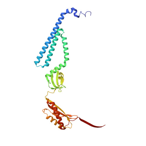
State-specific morphological deformations of the lipid bilayer explain mechanosensitive gating of MscS ion channels.
Park, Y.C., Reddy, B., Bavi, N., Perozo, E., Faraldo-Gomez, J.D.(2023) Elife 12
- PubMed: 36715097 Search on PubMedSearch on PubMed Central
- DOI: https://doi.org/10.7554/eLife.81445
- Primary Citation Related Structures:
8DDJ - PubMed Abstract:
The force-from-lipids hypothesis of cellular mechanosensation posits that membrane channels open and close in response to changes in the physical state of the lipid bilayer, induced for example by lateral tension. Here, we investigate the molecular basis for this transduction mechanism by studying the mechanosensitive ion channel MscS from Escherichia coli and its eukaryotic homolog MSL1 from Arabidopsis thaliana. First, we use single-particle cryo-electron microscopy to determine the structure of a novel open conformation of wild-type MscS, stabilized in a thinned lipid nanodisc. Compared with the closed state, the structure shows a reconfiguration of helices TM1, TM2, and TM3a, and widening of the central pore. Based on these structures, we examined how the morphology of the membrane is altered upon gating, using molecular dynamics simulations. The simulations reveal that closed-state MscS causes drastic protrusions in the inner leaflet of the lipid bilayer, both in the absence and presence of lateral tension, and for different lipid compositions. These deformations arise to provide adequate solvation to hydrophobic crevices under the TM1-TM2 hairpin, and clearly reflect a high-energy conformation for the membrane, particularly under tension. Strikingly, these protrusions are largely eradicated upon channel opening. An analogous computational study of open and closed MSL1 recapitulates these findings. The gating equilibrium of MscS channels thus appears to be dictated by opposing conformational preferences, namely those of the lipid membrane and of the protein structure. We propose a membrane deformation model of mechanosensation, which posits that tension shifts the gating equilibrium towards the conductive state not because it alters the mode in which channel and lipids interact, but because it increases the energetic cost of the morphological perturbations in the membrane required by the closed state.
- Theoretical Molecular Biophysics Laboratory, National Heart, Lung and Blood Institute, National Institutes of Health, Bethesda, United States.
Organizational Affiliation: