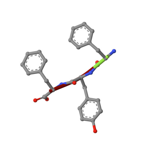
The rippled beta-sheet layer configuration-a novel supramolecular architecture based on predictions by Pauling and Corey.
Hazari, A., Sawaya, M.R., Vlahakis, N., Johnstone, T.C., Boyer, D., Rodriguez, J., Eisenberg, D., Raskatov, J.A.(2022) Chem Sci 13: 8947-8952
- PubMed: 36091211
- DOI: https://doi.org/10.1039/d2sc02531k
- Primary Citation of Related Structures:
8DDF, 8DDG, 8DDH - PubMed Abstract:
The rippled β-sheet is a peptidic structural motif related to but distinct from the pleated β-sheet. Both motifs were predicted in the 1950s by Pauling and Corey. The pleated β-sheet was since observed in countless proteins and peptides and is considered common textbook knowledge. Conversely, the rippled β-sheet only gained a meaningful experimental foundation in the past decade, and the first crystal structural study of rippled β-sheets was published as recently as this year. Noteworthy, the crystallized assembly stopped at the rippled β-dimer stage. It did not form the extended, periodic rippled β-sheet layer topography hypothesized by Pauling and Corey, thus calling the validity of their prediction into question. NMR work conducted since moreover shows that certain model peptides rather form pleated and not rippled β-sheets in solution. To determine whether the periodic rippled β-sheet layer configuration is viable, the field urgently needs crystal structures. Here we report on crystal structures of two racemic and one quasi-racemic aggregating peptide systems, all of which yield periodic rippled antiparallel β-sheet layers that are in excellent agreement with the predictions by Pauling and Corey. Our study establishes the rippled β-sheet layer configuration as a motif with general features and opens the road to structure-based design of unique supramolecular architectures.
- Dept. of Chemistry and Biochemistry, UCSC 1156 High Street Santa Cruz CA 95064 USA jraskato@ucsc.edu.
Organizational Affiliation:


