8DC0 | pdb_00008dc0

Rat Betaglycan Zona Pellucida Domain (ZPC) in complex with mini monomer TGFb2 (mmTGF-b2-7M2R)


Experimental Data Snapshot

  • Method: X-RAY DIFFRACTION
  • Resolution: 1.93 Å
  • R-Value Free: 
    0.257 (Depositor), 0.257 (DCC) 
  • R-Value Work: 
    0.219 (Depositor), 0.218 (DCC) 
  • R-Value Observed: 
    0.222 (Depositor) 

Starting Models: experimental
View more details

wwPDB Validation 3D Report Full Report

Validation slider image for 8DC0

This is version 1.3 of the entry. See complete history

Literature

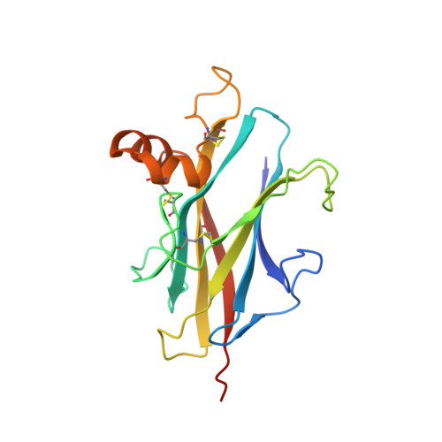
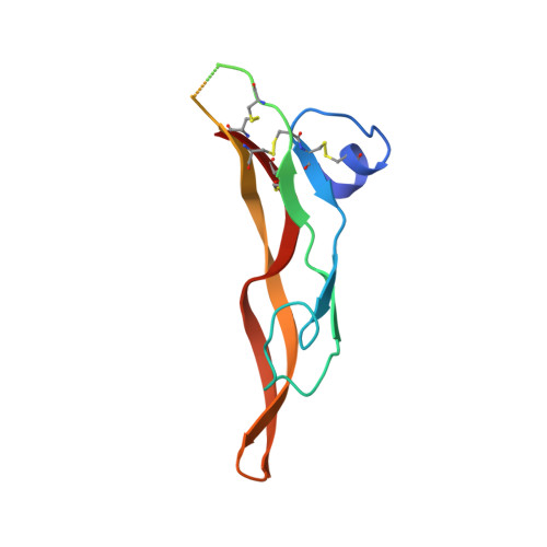
Structures of TGF-beta with betaglycan and signaling receptors reveal mechanisms of complex assembly and signaling.

Wieteska, L.Taylor, A.B.Punch, E.Coleman, J.A.Conway, I.O.Lin, Y.F.Byeon, C.H.Hinck, C.S.Krzysiak, T.Ishima, R.Lopez-Casillas, F.Cherepanov, P.Bernard, D.J.Hill, C.S.Hinck, A.P.

(2025) Nat Commun 16: 1778-1778

  • DOI: https://doi.org/10.1038/s41467-025-56796-9
  • Primary Citation Related Structures: 
    8DC0, 9B9F, 9FDY, 9FK5, 9FKP

  • PubMed Abstract: 

    Betaglycan (BG) is a transmembrane co-receptor of the transforming growth factor-β (TGF-β) family of signaling ligands. It is essential for embryonic development, tissue homeostasis and fertility in adults. It functions by enabling binding of the three TGF-β isoforms to their signaling receptors and is additionally required for inhibin A (InhA) activity. Despite its requirement for the functions of TGF-βs and InhA in vivo, structural information explaining BG ligand selectivity and its mechanism of action is lacking. Here, we determine the structure of TGF-β bound both to BG and the signaling receptors, TGFBR1 and TGFBR2. We identify key regions responsible for ligand engagement, which has revealed binding interfaces that differ from those described for the closely related co-receptor of the TGF-β family, endoglin, thus demonstrating remarkable evolutionary adaptation to enable ligand selectivity. Finally, we provide a structural explanation for the hand-off mechanism underlying TGF-β signal potentiation.


  • Organizational Affiliation
    • Department of Structural Biology, University of Pittsburgh, Pittsburgh, PA, USA.

Macromolecule Content 

  • Total Structure Weight: 32.86 kDa 
  • Atom Count: 2,131 
  • Modeled Residue Count: 255 
  • Deposited Residue Count: 286 
  • Unique protein chains: 2

Macromolecules

Find similar proteins by:|  3D Structure
Entity ID: 1
MoleculeChains  Sequence LengthOrganismDetailsImage
Transforming growth factor beta receptor type 3174Rattus norvegicusMutation(s): 0 
Gene Names: Tgfbr3
UniProt
Find proteins for P26342 (Rattus norvegicus)
Explore P26342 
Go to UniProtKB:  P26342
Entity Groups
Sequence Clusters30% Identity50% Identity70% Identity90% Identity95% Identity100% Identity
UniProt GroupP26342
Sequence Annotations
Expand
Reference Sequence
Find similar proteins by:|  3D Structure
Entity ID: 2
MoleculeChains  Sequence LengthOrganismDetailsImage
Transforming growth factor beta-2112Homo sapiensMutation(s): 0 
Gene Names: TGFB2
UniProt & NIH Common Fund Data Resources
Find proteins for P61812 (Homo sapiens)
Explore P61812 
Go to UniProtKB:  P61812
PHAROS:  P61812
GTEx:  ENSG00000092969 
Entity Groups
Sequence Clusters30% Identity50% Identity70% Identity90% Identity95% Identity100% Identity
UniProt GroupP61812
Sequence Annotations
Expand
Reference Sequence

Experimental Data & Validation

Experimental Data

  • Method: X-RAY DIFFRACTION
  • Resolution: 1.93 Å
  • R-Value Free:  0.257 (Depositor), 0.257 (DCC) 
  • R-Value Work:  0.219 (Depositor), 0.218 (DCC) 
  • R-Value Observed: 0.222 (Depositor) 
Space Group: I 2 2 2
Unit Cell:
Length ( Å )Angle ( ˚ )
a = 76.456α = 90
b = 90.453β = 90
c = 98.872γ = 90
Software Package:
Software NamePurpose
PHENIXrefinement
autoPROCdata reduction
Aimlessdata scaling
PHASERphasing

Structure Validation

View Full Validation Report


Entry History 

& Funding Information

Deposition Data


Funding OrganizationLocationGrant Number
National Institutes of Health/National Institute of General Medical Sciences (NIH/NIGMS)United StatesGM058067
H2020 Marie Curie Actions of the European CommissionEuropean Union893196

Revision History  (Full details and data files)

  • Version 1.0: 2023-06-21
    Type: Initial release
  • Version 1.1: 2023-10-25
    Changes: Data collection, Refinement description
  • Version 1.2: 2024-10-23
    Changes: Structure summary
  • Version 1.3: 2025-11-19
    Changes: Database references