8DBB | pdb_00008dbb

Crystal structure of DDT with the selective inhibitor 2,5-Pyridinedicarboxylic Acid


Experimental Data Snapshot

  • Method: X-RAY DIFFRACTION
  • Resolution: 1.30 Å
  • R-Value Free: 
    0.157 (Depositor), 0.156 (DCC) 
  • R-Value Work: 
    0.121 (Depositor), 0.121 (DCC) 
  • R-Value Observed: 
    0.123 (Depositor) 

Starting Model: experimental
View more details

wwPDB Validation 3D Report Full Report

Validation slider image for 8DBB

Ligand Structure Quality Assessment 


This is version 1.2 of the entry. See complete history

Literature

2,5-Pyridinedicarboxylic acid is a bioactive and highly selective inhibitor of D-dopachrome tautomerase.

Parkins, A.Das, P.Prahaladan, V.Rangel, V.M.Xue, L.Sankaran, B.Bhandari, V.Pantouris, G.

(2023) Structure 31: 355

  • DOI: https://doi.org/10.1016/j.str.2023.01.008
  • Primary Citation Related Structures: 
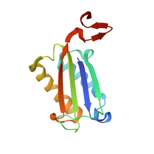
    8DBB

  • PubMed Abstract: 

    Macrophage migration inhibitory factor (MIF) and D-dopachrome tautomerase (D-DT) are two pleotropic cytokines, which are coexpressed in various cell types to activate the cell surface receptor CD74. Via the MIF/CD74 and D-DT/CD74 axes, the two proteins exhibit either beneficial or deleterious effect on human diseases. In this study, we report the identification of 2,5-pyridinedicarboxylic acid (a.k.a. 1) that effectively blocks the D-DT-induced activation of CD74 and demonstrates an impressive 79-fold selectivity for D-DT over MIF. Crystallographic characterization of D-DT-1 elucidates the binding features of 1 and reveals previously unrecognized differences between the MIF and D-DT active sites that explain the ligand's functional selectivity. The commercial availability, low cost, and high selectivity make 1 the ideal tool for studying the pathophysiological functionality of D-DT in disease models. At the same time, our comprehensive biochemical, computational, and crystallographic analyses serve as a guide for generating highly potent and selective D-DT inhibitors.


  • Organizational Affiliation
    • Department of Chemistry, University of the Pacific, Stockton, CA 95211, USA.

Macromolecule Content 

  • Total Structure Weight: 38.87 kDa 
  • Atom Count: 3,192 
  • Modeled Residue Count: 351 
  • Deposited Residue Count: 354 
  • Unique protein chains: 1

Macromolecules

Find similar proteins by:|  3D Structure
Entity ID: 1
MoleculeChains  Sequence LengthOrganismDetailsImage
D-dopachrome decarboxylase
A, B, C
118Homo sapiensMutation(s): 0 
Gene Names: DDT
EC: 4.1.1.84
UniProt & NIH Common Fund Data Resources
Find proteins for P30046 (Homo sapiens)
Explore P30046 
Go to UniProtKB:  P30046
PHAROS:  P30046
GTEx:  ENSG00000099977 
Entity Groups
Sequence Clusters30% Identity50% Identity70% Identity90% Identity95% Identity100% Identity
UniProt GroupP30046
Sequence Annotations
Expand
Reference Sequence

Experimental Data & Validation

Experimental Data

  • Method: X-RAY DIFFRACTION
  • Resolution: 1.30 Å
  • R-Value Free:  0.157 (Depositor), 0.156 (DCC) 
  • R-Value Work:  0.121 (Depositor), 0.121 (DCC) 
  • R-Value Observed: 0.123 (Depositor) 
Space Group: P 3
Unit Cell:
Length ( Å )Angle ( ˚ )
a = 83.721α = 90
b = 83.721β = 90
c = 40.633γ = 120
Software Package:
Software NamePurpose
XDSdata reduction
Aimlessdata scaling
PHASERphasing
REFMACrefinement
PDB_EXTRACTdata extraction

Structure Validation

View Full Validation Report


Ligand Structure Quality Assessment 


Entry History 

& Funding Information

Deposition Data


Funding OrganizationLocationGrant Number
Other privateUnited States--

Revision History  (Full details and data files)

  • Version 1.0: 2023-03-08
    Type: Initial release
  • Version 1.1: 2023-03-15
    Changes: Database references
  • Version 1.2: 2023-10-25
    Changes: Data collection, Refinement description