8D3H | pdb_00008d3h

Crystal structure of human Apoptosis-Inducing Factor (AIF) W196A mutant complexed with 7-chloroquinolin-4-amine


Experimental Data Snapshot

  • Method: X-RAY DIFFRACTION
  • Resolution: 2.51 Å
  • R-Value Free: 
    0.231 (Depositor), 0.234 (DCC) 
  • R-Value Work: 
    0.204 (Depositor), 0.207 (DCC) 
  • R-Value Observed: 
    0.206 (Depositor) 

Starting Model: experimental
View more details

wwPDB Validation 3D Report Full Report

Validation slider image for 8D3H

Ligand Structure Quality Assessment 


This is version 1.2 of the entry. See complete history

Literature

Chemical screening by time-resolved X-ray scattering to discover allosteric probes.

Brosey, C.A.Link, T.M.Shen, R.Moiani, D.Burnett, K.Hura, G.L.Jones, D.E.Tainer, J.A.

(2024) Nat Chem Biol 

  • DOI: https://doi.org/10.1038/s41589-024-01609-1
  • Primary Citation Related Structures: 
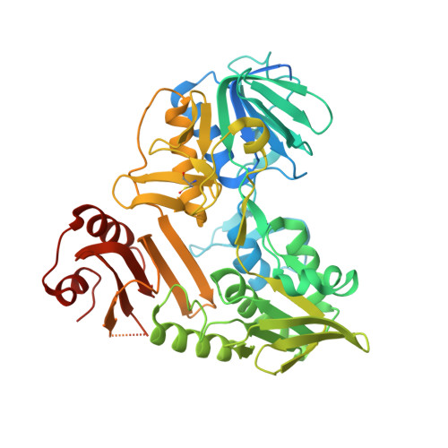
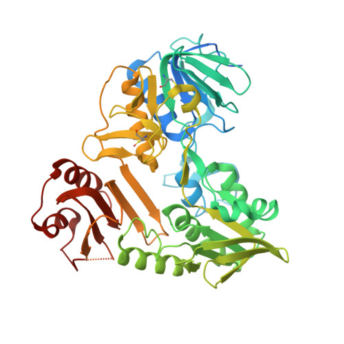
    8D3E, 8D3G, 8D3H, 8D3I, 8D3J, 8D3K, 8D3N, 8D3O

  • PubMed Abstract: 

    Drug discovery relies on efficient identification of small-molecule leads and their interactions with macromolecular targets. However, understanding how chemotypes impact mechanistically important conformational states often remains secondary among high-throughput discovery methods. Here, we present a conformational discovery pipeline integrating time-resolved, high-throughput small-angle X-ray scattering (TR-HT-SAXS) and classic fragment screening applied to allosteric states of the mitochondrial import oxidoreductase apoptosis-inducing factor (AIF). By monitoring oxidized and X-ray-reduced AIF states, TR-HT-SAXS leverages structure and kinetics to generate a multidimensional screening dataset that identifies fragment chemotypes allosterically stimulating AIF dimerization. Fragment-induced dimerization rates, quantified with time-resolved SAXS similarity analysis (k VR ), capture structure-activity relationships (SAR) across the top-ranked 4-aminoquinoline chemotype. Crystallized AIF-aminoquinoline complexes validate TR-SAXS-guided SAR, supporting this conformational chemotype for optimization. AIF-aminoquinoline structures and mutational analysis reveal active site F482 as an underappreciated allosteric stabilizer of AIF dimerization. This conformational discovery pipeline illustrates TR-HT-SAXS as an effective technology for targeting chemical leads to important macromolecular states.


  • Organizational Affiliation
    • Department of Molecular and Cellular Oncology, The University of Texas MD Anderson Cancer Center, Houston, TX, USA. cabrosey@mdanderson.org.

Macromolecule Content 

  • Total Structure Weight: 120.75 kDa 
  • Atom Count: 6,802 
  • Modeled Residue Count: 866 
  • Deposited Residue Count: 1,086 
  • Unique protein chains: 2

Macromolecules

Find similar proteins by:|  3D Structure
Entity ID: 1
MoleculeChains  Sequence LengthOrganismDetailsImage
Apoptosis-inducing factor 1, mitochondrial543Homo sapiensMutation(s): 1 
Gene Names: AIFM1AIFPDCD8
EC: 1.6.99
UniProt & NIH Common Fund Data Resources
Find proteins for O95831 (Homo sapiens)
Explore O95831 
Go to UniProtKB:  O95831
PHAROS:  O95831
GTEx:  ENSG00000156709 
Entity Groups
Sequence Clusters30% Identity50% Identity70% Identity90% Identity95% Identity100% Identity
UniProt GroupO95831
Sequence Annotations
Expand
Reference Sequence
Find similar proteins by:|  3D Structure
Entity ID: 2
MoleculeChains  Sequence LengthOrganismDetailsImage
Apoptosis-inducing factor 1, mitochondrial543Homo sapiensMutation(s): 1 
Gene Names: AIFM1AIFPDCD8
EC: 1.6.99
UniProt & NIH Common Fund Data Resources
Find proteins for O95831 (Homo sapiens)
Explore O95831 
Go to UniProtKB:  O95831
PHAROS:  O95831
GTEx:  ENSG00000156709 
Entity Groups
Sequence Clusters30% Identity50% Identity70% Identity90% Identity95% Identity100% Identity
UniProt GroupO95831
Sequence Annotations
Expand
Reference Sequence

Experimental Data & Validation

Experimental Data

  • Method: X-RAY DIFFRACTION
  • Resolution: 2.51 Å
  • R-Value Free:  0.231 (Depositor), 0.234 (DCC) 
  • R-Value Work:  0.204 (Depositor), 0.207 (DCC) 
  • R-Value Observed: 0.206 (Depositor) 
Space Group: P 21 21 21
Unit Cell:
Length ( Å )Angle ( ˚ )
a = 90.787α = 90
b = 114.767β = 90
c = 120.907γ = 90
Software Package:
Software NamePurpose
XDSdata reduction
Aimlessdata scaling
PHASERphasing
PHENIXrefinement
Cootmodel building

Structure Validation

View Full Validation Report


Ligand Structure Quality Assessment 


Entry History 

& Funding Information

Deposition Data


Funding OrganizationLocationGrant Number
National Institutes of Health/National Cancer Institute (NIH/NCI)United StatesR35 CA220430
National Institutes of Health/National Cancer Institute (NIH/NCI)United StatesP01 CA092584
Cancer Prevention and Research Institute of Texas (CPRIT)United StatesRP180813

Revision History  (Full details and data files)

  • Version 1.0: 2023-11-08
    Type: Initial release
  • Version 1.1: 2024-05-29
    Changes: Database references
  • Version 1.2: 2026-03-04
    Changes: Refinement description, Structure summary