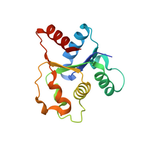
8D0E

Human SARM1 TIR domain bound to NB-7


Experimental Data Snapshot

  • Method: X-RAY DIFFRACTION
  • Resolution: 1.88 Å
  • R-Value Free: 0.228 
  • R-Value Work: 0.183 
  • R-Value Observed: 0.186 

Starting Model: experimental
View more details

wwPDB Validation   3D Report Full Report


Ligand Structure Quality Assessment 


This is version 1.2 of the entry. See complete history


Literature

Uncompetitive, adduct-forming SARM1 inhibitors are neuroprotective in preclinical models of nerve injury and disease.

Bratkowski, M.Burdett, T.C.Danao, J.Wang, X.Mathur, P.Gu, W.Beckstead, J.A.Talreja, S.Yang, Y.S.Danko, G.Park, J.H.Walton, M.Brown, S.P.Tegley, C.M.Joseph, P.R.B.Reynolds, C.H.Sambashivan, S.

(2022) Neuron 110: 3711

  • DOI: https://doi.org/10.1016/j.neuron.2022.08.017
  • Primary Citation of Related Structures:  
    8D0C, 8D0D, 8D0E, 8D0F, 8D0G, 8D0H, 8D0I, 8D0J, 8D0M

  • PubMed Abstract: 

    Axon degeneration is an early pathological event in many neurological diseases. The identification of the nicotinamide adenine dinucleotide (NAD) hydrolase SARM1 as a central metabolic sensor and axon executioner presents an exciting opportunity to develop novel neuroprotective therapies that can prevent or halt the degenerative process, yet limited progress has been made on advancing efficacious inhibitors. We describe a class of NAD-dependent active-site SARM1 inhibitors that function by intercepting NAD hydrolysis and undergoing covalent conjugation with the reaction product adenosine diphosphate ribose (ADPR). The resulting small-molecule ADPR adducts are highly potent and confer compelling neuroprotection in preclinical models of neurological injury and disease, validating this mode of inhibition as a viable therapeutic strategy. Additionally, we show that the most potent inhibitor of CD38, a related NAD hydrolase, also functions by the same mechanism, further underscoring the broader applicability of this mechanism in developing therapies against this class of enzymes.


  • Organizational Affiliation

    Biology Department, Nura Bio Inc., South San Francisco, CA 94080, USA.


Macromolecules
Find similar proteins by:  (by identity cutoff)  |  3D Structure
Entity ID: 1
MoleculeChains Sequence LengthOrganismDetailsImage
NAD(+) hydrolase SARM1
A, B
143Homo sapiensMutation(s): 0 
Gene Names: SARM1KIAA0524SAMD2SARM
EC: 3.2.2.6 (PDB Primary Data), 3.2.2 (PDB Primary Data)
UniProt & NIH Common Fund Data Resources
Find proteins for Q6SZW1 (Homo sapiens)
Explore Q6SZW1 
Go to UniProtKB:  Q6SZW1
PHAROS:  Q6SZW1
GTEx:  ENSG00000004139 
Entity Groups  
Sequence Clusters30% Identity50% Identity70% Identity90% Identity95% Identity100% Identity
UniProt GroupQ6SZW1
Sequence Annotations
Expand
  • Reference Sequence
Small Molecules
Ligands 1 Unique
IDChains Name / Formula / InChI Key2D Diagram3D Interactions
Q0C
Query on Q0C

Download Ideal Coordinates CCD File 
C [auth A],
D [auth B]
3-(4-chlorophenyl)-N-[4-methyl-3-(pyridin-4-yl)-1H-pyrazol-5-yl]propanamide
C18 H17 Cl N4 O
YPBDFUFBFVQJPF-UHFFFAOYSA-N
Experimental Data & Validation

Experimental Data

  • Method: X-RAY DIFFRACTION
  • Resolution: 1.88 Å
  • R-Value Free: 0.228 
  • R-Value Work: 0.183 
  • R-Value Observed: 0.186 
  • Space Group: P 2 21 21
Unit Cell:
Length ( Å )Angle ( ˚ )
a = 32.927α = 90
b = 87.232β = 90
c = 116.634γ = 90
Software Package:
Software NamePurpose
PHENIXrefinement
XDSdata reduction
Aimlessdata scaling
PHASERphasing

Structure Validation

View Full Validation Report


Ligand Structure Quality Assessment 


Entry History & Funding Information

Deposition Data


Funding OrganizationLocationGrant Number
Other privateUnited States--

Revision History  (Full details and data files)

  • Version 1.0: 2022-09-21
    Type: Initial release
  • Version 1.1: 2022-11-30
    Changes: Database references
  • Version 1.2: 2023-10-25
    Changes: Data collection, Refinement description