The Cryo-EM STRUCTURE of Renal Amyloid Fibril Suggests Structurally Homogeneous Multiorgan Aggregation in AL Amyloidosis.
Puri, S., Schulte, T., Chaves-Sanjuan, A., Mazzini, G., Caminito, S., Pappone, C., Anastasia, L., Milani, P., Merlini, G., Bolognesi, M., Nuvolone, M., Palladini, G., Ricagno, S.(2023) J Mol Biology 435: 168215-168215
- PubMed: 37516426 Search on PubMed
- DOI: https://doi.org/10.1016/j.jmb.2023.168215
- Primary Citation Related Structures:
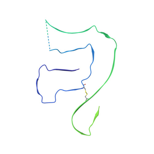
8CPE - PubMed Abstract:
Immunoglobulin light chain amyloidosis (AL) is caused by the aberrant production of amyloidogenic light chains (LC) that accumulate as amyloid deposits in vital organs. Distinct LC sequences in each patient yield distinct amyloid structures. However different tissue microenvironments may also cause identical protein precursors to adopt distinct amyloid structures. To address the impact of the tissue environment on the structural polymorphism of amyloids, we extracted fibrils from the kidney of an AL patient (AL55) whose cardiac amyloid structure was previously determined by our group. Here we show that the 4.0 Å resolution cryo-EM structure of the renal fibril is virtually identical to that reported for the cardiac fibril. These results provide the first structural evidence that LC amyloids independently deposited in different organs of the same AL patient share a common fold.
- Department of Biosciences, Università degli Studi di Milano, Milan, Italy. Electronic address: https://twitter.com/@Saritapuri1504.
Organizational Affiliation: