8COK | pdb_00008cok

Structural analysis of ING3 protein and its binding to histone H3


Experimental Data Snapshot

  • Method: X-RAY DIFFRACTION
  • Resolution: 2.91 Å
  • R-Value Free: 
    0.315 (Depositor), 0.316 (DCC) 
  • R-Value Work: 
    0.270 (Depositor), 0.270 (DCC) 
  • R-Value Observed: 
    0.275 (Depositor) 

Starting Model: experimental
View more details

wwPDB Validation 3D Report Full Report

Validation slider image for 8COK

This is version 1.2 of the entry. See complete history

Literature

Structural analysis of ING3 protein and histone H3 binding.

Ferreras-Gutierrez, M.Chaves-Arquero, B.Gonzalez-Magana, A.Merino, N.Amusategui-Mateu, I.Huecas, S.Medrano, F.J.Blanco, F.J.

(2023) Int J Biol Macromol 242: 124724-124724

  • DOI: https://doi.org/10.1016/j.ijbiomac.2023.124724
  • Primary Citation Related Structures: 
    7ZMX, 8COK

  • PubMed Abstract: 

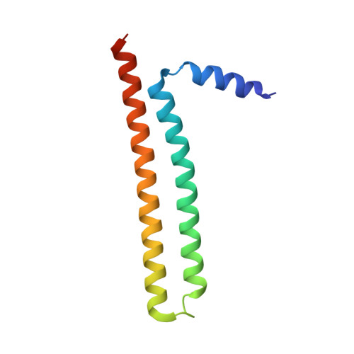
    Proteins belonging to the ING family regulate the transcriptional state of chromatin by recruiting remodeling complexes to sites with histone H3 trimethylated at Lysine 4 (H3K4me3). This modification is recognized by the Plant HomeoDomain (PHD) present at the C-terminal region of the five ING proteins. ING3 facilitates acetylation of histones H2A and H4 by the NuA4-Tip60 MYST histone acetyl transferase complex, and it has been proposed to be an oncoprotein. The crystal structure of the N-terminal domain of ING3 shows that it forms homodimers with an antiparallel coiled-coil fold. The crystal structure of the PHD is similar to those of its four homologs. These structures explain the possible deleterious effects of ING3 mutations detected in tumors. The PHD binds histone H3K4me3 with low-micromolar, and binds the non-methylated histone with a 54-fold reduced affinity. Our structure explains the impact of site directed mutagenesis experiments on histone recognition. These structural features could not be confirmed for the full-length protein as solubility was insufficient for structural studies, but the structure of its folded domains suggest a conserved structural organization for the ING proteins as homodimers and bivalent readers of the histone H3K4me3 mark.


  • Organizational Affiliation
    • Centro de Investigaciones Biológicas Margarita Salas (CIB), CSIC, Madrid 28040, Spain.

Macromolecule Content 

  • Total Structure Weight: 25.28 kDa 
  • Atom Count: 1,706 
  • Modeled Residue Count: 199 
  • Deposited Residue Count: 208 
  • Unique protein chains: 1

Macromolecules

Find similar proteins by:|  3D Structure
Entity ID: 1
MoleculeChains  Sequence LengthOrganismDetailsImage
Inhibitor of growth protein 3
A, B
104Homo sapiensMutation(s): 0 
Gene Names: Ing3
UniProt & NIH Common Fund Data Resources
Find proteins for Q9NXR8 (Homo sapiens)
Explore Q9NXR8 
Go to UniProtKB:  Q9NXR8
PHAROS:  Q9NXR8
GTEx:  ENSG00000071243 
Entity Groups
Sequence Clusters30% Identity50% Identity70% Identity90% Identity95% Identity100% Identity
UniProt GroupQ9NXR8
Sequence Annotations
Expand
Reference Sequence

Experimental Data & Validation

Experimental Data

  • Method: X-RAY DIFFRACTION
  • Resolution: 2.91 Å
  • R-Value Free:  0.315 (Depositor), 0.316 (DCC) 
  • R-Value Work:  0.270 (Depositor), 0.270 (DCC) 
  • R-Value Observed: 0.275 (Depositor) 
Space Group: I 4 3 2
Unit Cell:
Length ( Å )Angle ( ˚ )
a = 149.394α = 90
b = 149.394β = 90
c = 149.394γ = 90
Software Package:
Software NamePurpose
PHENIXrefinement
PHENIXrefinement
autoPROCdata reduction
autoPROCdata scaling
MrBUMPphasing

Structure Validation

View Full Validation Report


Entry History 

& Funding Information

Deposition Data


Funding OrganizationLocationGrant Number
Ministerio de Ciencia e Innovacion (MCIN)SpainPID2020-113225GB-I00

Revision History  (Full details and data files)

  • Version 1.0: 2023-05-17
    Type: Initial release
  • Version 1.1: 2023-05-24
    Changes: Database references
  • Version 1.2: 2023-09-20
    Changes: Data collection, Refinement description