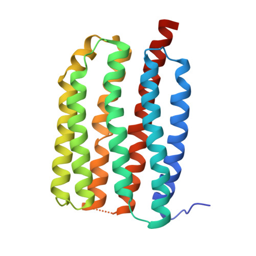
Structural insights into the mechanism and dynamics of proteorhodopsin biogenesis and retinal scavenging.
Hirschi, S., Lemmin, T., Ayoub, N., Kalbermatter, D., Pellegata, D., Ucurum, Z., Gertsch, J., Fotiadis, D.(2024) Nat Commun 15: 6950-6950
- PubMed: 39138159
- DOI: https://doi.org/10.1038/s41467-024-50960-3
- Primary Citation of Related Structures:
8CNK, 8CQC, 8CQD - PubMed Abstract:
Microbial ion-pumping rhodopsins (MRs) are extensively studied retinal-binding membrane proteins. However, their biogenesis, including oligomerisation and retinal incorporation, remains poorly understood. The bacterial green-light absorbing proton pump proteorhodopsin (GPR) has emerged as a model protein for MRs and is used here to address these open questions using cryo-electron microscopy (cryo-EM) and molecular dynamics (MD) simulations. Specifically, conflicting studies regarding GPR stoichiometry reported pentamer and hexamer mixtures without providing possible assembly mechanisms. We report the pentameric and hexameric cryo-EM structures of a GPR mutant, uncovering the role of the unprocessed N-terminal signal peptide in the assembly of hexameric GPR. Furthermore, certain proteorhodopsin-expressing bacteria lack retinal biosynthesis pathways, suggesting that they scavenge the cofactor from their environment. We shed light on this hypothesis by solving the cryo-EM structure of retinal-free proteoopsin, which together with mass spectrometry and MD simulations suggests that decanoate serves as a temporary placeholder for retinal in the chromophore binding pocket. Further MD simulations elucidate possible pathways for the exchange of decanoate and retinal, offering a mechanism for retinal scavenging. Collectively, our findings provide insights into the biogenesis of MRs, including their oligomeric assembly, variations in protomer stoichiometry and retinal incorporation through a potential cofactor scavenging mechanism.
Organizational Affiliation:
Institute of Biochemistry and Molecular Medicine, University of Bern, 3012, Bern, Switzerland. stephan.hirschi@bioch.ox.ac.uk.


