8CKZ

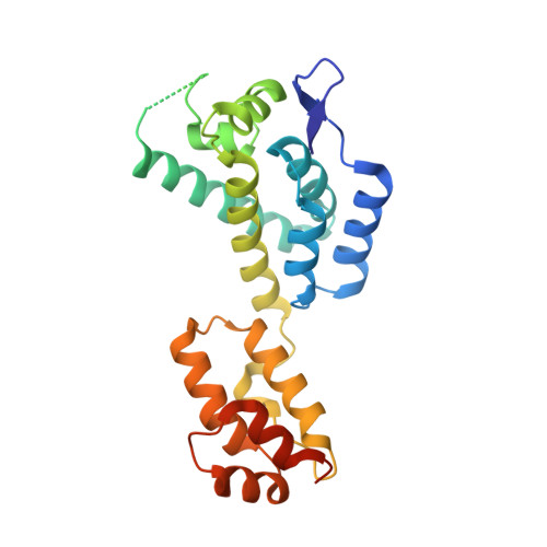
HIV-1 mature capsid pentamer from CA-IP6 CLPs bound to Nup153 peptide


Experimental Data Snapshot

  • Method: ELECTRON MICROSCOPY
  • Resolution: 3.07 Å
  • Aggregation State: PARTICLE 
  • Reconstruction Method: SINGLE PARTICLE 

Starting Model: experimental
View more details

wwPDB Validation   3D Report Full Report


This is version 1.1 of the entry. See complete history


Literature

Two structural switches in HIV-1 capsid regulate capsid curvature and host factor binding.

Stacey, J.C.V.Tan, A.Lu, J.M.James, L.C.Dick, R.A.Briggs, J.A.G.

(2023) Proc Natl Acad Sci U S A 120: e2220557120-e2220557120

  • DOI: https://doi.org/10.1073/pnas.2220557120
  • Primary Citation of Related Structures:  
    8CKV, 8CKW, 8CKX, 8CKY, 8CKZ, 8CL0, 8CL1, 8CL2, 8CL3, 8CL4

  • PubMed Abstract: 

    The mature HIV-1 capsid protects the viral genome and interacts with host proteins to travel from the cell periphery into the nucleus. To achieve this, the capsid protein, CA, constructs conical capsids from a lattice of hexamers and pentamers, and engages in and then relinquishes multiple interactions with cellular proteins in an orchestrated fashion. Cellular host factors including Nup153, CPSF6, and Sec24C engage the same pocket within CA hexamers. How CA assembles pentamers and hexamers of different curvatures, how CA oligomerization states or curvature might modulate host-protein interactions, and how binding of multiple cofactors to a single site is coordinated, all remain to be elucidated. Here, using single-particle cryoEM, we have determined the structure of the mature HIV-1 CA pentamer and hexamer from conical CA-IP 6 polyhedra to ~3 Å resolution. We also determined structures of hexamers in the context of multiple lattice curvatures and number of pentamer contacts. Comparison of these structures, bound or not to host protein peptides, revealed two structural switches within HIV-1 CA that modulate peptide binding according to CA lattice curvature and whether CA is hexameric or pentameric. These observations suggest that the conical HIV-1 capsid has different host-protein binding properties at different positions on its surface, which may facilitate cell entry and represent an evolutionary advantage of conical morphology.


  • Organizational Affiliation

    Department of Cell and Virus Structure, Max Planck Institute of Biochemistry, Martinsried 82512, Germany.


Macromolecules
Find similar proteins by:  (by identity cutoff)  |  3D Structure
Entity ID: 1
MoleculeChains Sequence LengthOrganismDetailsImage
Gag polyprotein
A, B, C
232Human immunodeficiency virus 1Mutation(s): 0 
Gene Names: gag
UniProt
Find proteins for P12493 (Human immunodeficiency virus type 1 group M subtype B (isolate NY5))
Explore P12493 
Go to UniProtKB:  P12493
Entity Groups  
Sequence Clusters30% Identity50% Identity70% Identity90% Identity95% Identity100% Identity
UniProt GroupP12493
Sequence Annotations
Expand
  • Reference Sequence
Experimental Data & Validation

Experimental Data

  • Method: ELECTRON MICROSCOPY
  • Resolution: 3.07 Å
  • Aggregation State: PARTICLE 
  • Reconstruction Method: SINGLE PARTICLE 
EM Software:
TaskSoftware PackageVersion
MODEL REFINEMENTISOLDE
MODEL REFINEMENTCoot
MODEL REFINEMENTPHENIX
RECONSTRUCTIONcryoSPARC3.3.2

Structure Validation

View Full Validation Report


Entry History & Funding Information

Deposition Data


Funding OrganizationLocationGrant Number
Max Planck SocietyGermany--
National Institutes of Health/National Institute Of Allergy and Infectious Diseases (NIH/NIAID)United StatesR01AI147890
National Institutes of Health/National Institute Of Allergy and Infectious Diseases (NIH/NIAID)United States5U54AI150472-09
National Institutes of Health/National Institute Of Allergy and Infectious Diseases (NIH/NIAID)United StatesU54 AI170855-01
Medical Research Council (MRC, United Kingdom)United KingdomMC_UP_1201/16

Revision History  (Full details and data files)

  • Version 1.0: 2023-04-26
    Type: Initial release
  • Version 1.1: 2024-07-24
    Changes: Data collection, Refinement description