8CKK | pdb_00008ckk

Semaphorin-5A TSR 3-4 domains in complex with nitrate


Experimental Data Snapshot

  • Method: X-RAY DIFFRACTION
  • Resolution: 1.56 Å
  • R-Value Free: 
    0.229 (Depositor), 0.228 (DCC) 
  • R-Value Work: 
    0.197 (Depositor), 0.196 (DCC) 
  • R-Value Observed: 
    0.198 (Depositor) 

Starting Model: experimental
View more details

wwPDB Validation 3D Report Full Report

Validation slider image for 8CKK

Ligand Structure Quality Assessment 


This is version 1.2 of the entry. See complete history

Literature

Structure and function of Semaphorin-5A glycosaminoglycan interactions.

Nagy, G.N.Zhao, X.F.Karlsson, R.Wang, K.Duman, R.Harlos, K.El Omari, K.Wagner, A.Clausen, H.Miller, R.L.Giger, R.J.Jones, E.Y.

(2024) Nat Commun 15: 2723-2723

  • DOI: https://doi.org/10.1038/s41467-024-46725-7
  • Primary Citation Related Structures: 
    8CKG, 8CKK, 8CKL, 8CKM

  • PubMed Abstract: 

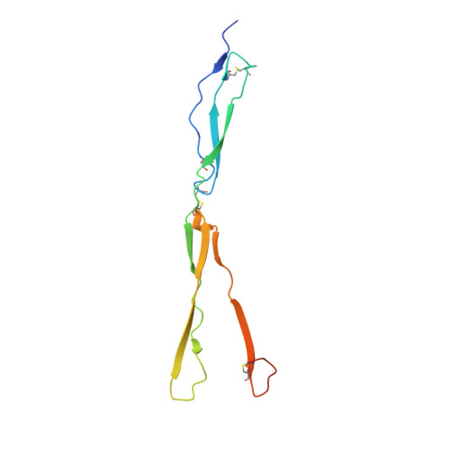
    Integration of extracellular signals by neurons is pivotal for brain development, plasticity, and repair. Axon guidance relies on receptor-ligand interactions crosstalking with extracellular matrix components. Semaphorin-5A (Sema5A) is a bifunctional guidance cue exerting attractive and inhibitory effects on neuronal growth through the interaction with heparan sulfate (HS) and chondroitin sulfate (CS) glycosaminoglycans (GAGs), respectively. Sema5A harbors seven thrombospondin type-1 repeats (TSR1-7) important for GAG binding, however the underlying molecular basis and functions in vivo remain enigmatic. Here we dissect the structural basis for Sema5A:GAG specificity and demonstrate the functional significance of this interaction in vivo. Using x-ray crystallography, we reveal a dimeric fold variation for TSR4 that accommodates GAG interactions. TSR4 co-crystal structures identify binding residues validated by site-directed mutagenesis. In vitro and cell-based assays uncover specific GAG epitopes necessary for TSR association. We demonstrate that HS-GAG binding is preferred over CS-GAG and mediates Sema5A oligomerization. In vivo, Sema5A:GAG interactions are necessary for Sema5A function and regulate Plexin-A2 dependent dentate progenitor cell migration. Our study rationalizes Sema5A associated developmental and neurological disorders and provides mechanistic insights into how multifaceted guidance functions of a single transmembrane cue are regulated by proteoglycans.


  • Organizational Affiliation
    • Division of Structural Biology, Wellcome Centre for Human Genetics, University of Oxford, Oxford, UK. nagy.gergely.nandor@vbk.bme.hu.

Macromolecule Content 

  • Total Structure Weight: 29.88 kDa 
  • Atom Count: 2,098 
  • Modeled Residue Count: 234 
  • Deposited Residue Count: 250 
  • Unique protein chains: 1

Macromolecules

Find similar proteins by:|  3D Structure
Entity ID: 1
MoleculeChains  Sequence LengthOrganismDetailsImage
Semaphorin-5A
A, B
125Homo sapiensMutation(s): 0 
Gene Names: SEMA5ASEMAF
UniProt & NIH Common Fund Data Resources
Find proteins for Q13591 (Homo sapiens)
Explore Q13591 
Go to UniProtKB:  Q13591
PHAROS:  Q13591
GTEx:  ENSG00000112902 
Entity Groups
Sequence Clusters30% Identity50% Identity70% Identity90% Identity95% Identity100% Identity
UniProt GroupQ13591
Glycosylation
Glycosylation Sites: 2Go to GlyGen: Q13591-1
Sequence Annotations
Expand
Reference Sequence

Small Molecules

Ligands 2 Unique
IDChains Name / Formula / InChI Key2D Diagram3D Interactions
MAN
(Subject of Investigation/LOI)

Query on MAN


Download:Ideal Coordinates CCD File
C [auth A],
M [auth A],
V [auth B],
W [auth B]
alpha-D-mannopyranose
C6 H12 O6
WQZGKKKJIJFFOK-PQMKYFCFSA-N
NO3

Query on NO3


Download:Ideal Coordinates CCD File
D [auth A]
E [auth A]
F [auth A]
G [auth A]
H [auth A]
D [auth A],
E [auth A],
F [auth A],
G [auth A],
H [auth A],
I [auth A],
J [auth A],
K [auth A],
L [auth A],
N [auth B],
O [auth B],
P [auth B],
Q [auth B],
R [auth B],
S [auth B],
T [auth B],
U [auth B]
NITRATE ION
N O3
NHNBFGGVMKEFGY-UHFFFAOYSA-N

Experimental Data & Validation

Experimental Data

  • Method: X-RAY DIFFRACTION
  • Resolution: 1.56 Å
  • R-Value Free:  0.229 (Depositor), 0.228 (DCC) 
  • R-Value Work:  0.197 (Depositor), 0.196 (DCC) 
  • R-Value Observed: 0.198 (Depositor) 
Space Group: P 21 21 21
Unit Cell:
Length ( Å )Angle ( ˚ )
a = 51.894α = 90
b = 68.347β = 90
c = 85.502γ = 90
Software Package:
Software NamePurpose
autoPROCdata processing
PHENIXrefinement
Cootmodel building
XDSdata reduction
Aimlessdata scaling
PHASERphasing

Structure Validation

View Full Validation Report


Ligand Structure Quality Assessment 


Entry History 

& Funding Information

Deposition Data


Funding OrganizationLocationGrant Number
Medical Research Council (MRC, United Kingdom)United KingdomMR/T000503/1

Revision History  (Full details and data files)

  • Version 1.0: 2024-02-28
    Type: Initial release
  • Version 1.1: 2024-09-11
    Changes: Database references
  • Version 1.2: 2024-11-13
    Changes: Structure summary