8CGE

The crystal structure of a cobalt-bound scFv reveals a Tetrameric polyHistidine motif (TetrHis)


Experimental Data Snapshot

  • Method: X-RAY DIFFRACTION
  • Resolution: 2.50 Å
  • R-Value Free: 0.248 
  • R-Value Work: 0.190 
  • R-Value Observed: 0.193 

wwPDB Validation   3D Report Full Report


This is version 1.0 of the entry. See complete history


Literature

Structure, dynamics and transferability of the metal-dependent polyhistidine tetramerization motif TetrHis for single-chain Fv antibodies.

Healey, R.D.Couillaud, L.Hoh, F.Mouhand, A.Fouillen, A.Couvineau, P.Granier, S.Leyrat, C.

(2023) Commun Chem 6: 160-160

  • DOI: https://doi.org/10.1038/s42004-023-00962-x
  • Primary Citation of Related Structures:  
    8CGE

  • PubMed Abstract: 

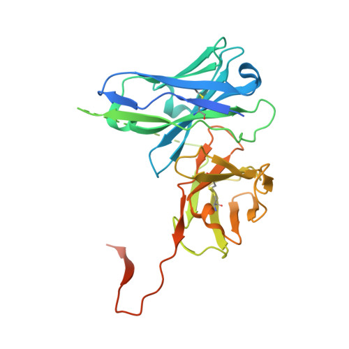
    The polyhistidine (6XHis) motif is one of the most ubiquitous protein purification tags. The 6XHis motif enables the binding of tagged proteins to various metals, which can be advantageously used for purification with immobilized metal affinity chromatography. Despite its popularity, protein structures encompassing metal-bound 6XHis are rare. Here, we obtained a 2.5 Å resolution crystal structure of a single chain Fv antibody (scFv) bearing a C-terminal sortase motif, 6XHis and TwinStrep tags (LPETGHHHHHHWSHPQFEK[G 3 S] 3 WSHPQFEK). The structure, obtained in the presence of cobalt, reveals a unique tetramerization motif (TetrHis) stabilized by 8 Co 2+ ions. The TetrHis motif contains four 6 residues-long β-strands, and each metal center coordinates 3 to 5 residues, including all 6XHis histidines. By combining dynamic light scattering, small angle x-ray scattering and molecular dynamics simulations, We investigated the influence of Co 2+ on the conformational dynamics of scFv 2A2, observing an open/close equilibrium of the monomer and the formation of cobalt-stabilized tetramers. By using a similar scFv design, we demonstrate the transferability of the tetramerization property. This novel metal-dependent tetramerization motif might be used as a fiducial marker for cryoelectron microscopy of scFv complexes, or even provide a starting point for designing metal-loaded biomaterials.


  • Organizational Affiliation

    IGF, University of Montpellier, CNRS, INSERM, Montpellier, France.


Macromolecules
Find similar proteins by:  (by identity cutoff)  |  3D Structure
Entity ID: 1
MoleculeChains Sequence LengthOrganismDetailsImage
Single Chain Fv 2A2
A, B
304synthetic constructMutation(s): 0 
Entity Groups  
Sequence Clusters30% Identity50% Identity70% Identity90% Identity95% Identity100% Identity
Sequence Annotations
Expand
  • Reference Sequence
Small Molecules
Ligands 3 Unique
IDChains Name / Formula / InChI Key2D Diagram3D Interactions
CXS
Query on CXS

Download Ideal Coordinates CCD File 
M [auth B]3-CYCLOHEXYL-1-PROPYLSULFONIC ACID
C9 H19 N O3 S
PJWWRFATQTVXHA-UHFFFAOYSA-N
SO4
Query on SO4

Download Ideal Coordinates CCD File 
E [auth A]
F [auth A]
G [auth A]
H [auth A]
I [auth A]
E [auth A],
F [auth A],
G [auth A],
H [auth A],
I [auth A],
J [auth A],
K [auth A],
L [auth A],
P [auth B],
Q [auth B],
R [auth B],
S [auth B],
T [auth B],
U [auth B],
V [auth B]
SULFATE ION
O4 S
QAOWNCQODCNURD-UHFFFAOYSA-L
CO (Subject of Investigation/LOI)
Query on CO

Download Ideal Coordinates CCD File 
C [auth A],
D [auth A],
N [auth B],
O [auth B]
COBALT (II) ION
Co
XLJKHNWPARRRJB-UHFFFAOYSA-N
Experimental Data & Validation

Experimental Data

  • Method: X-RAY DIFFRACTION
  • Resolution: 2.50 Å
  • R-Value Free: 0.248 
  • R-Value Work: 0.190 
  • R-Value Observed: 0.193 
  • Space Group: I 1 2 1
Unit Cell:
Length ( Å )Angle ( ˚ )
a = 97.496α = 90
b = 81.817β = 111.14
c = 111.045γ = 90
Software Package:
Software NamePurpose
BUSTERrefinement
PHENIXrefinement
xia2data reduction
xia2data scaling
PHASERphasing

Structure Validation

View Full Validation Report


Entry History & Funding Information

Deposition Data


Funding OrganizationLocationGrant Number
Not fundedFrance--

Revision History  (Full details and data files)

  • Version 1.0: 2023-08-09
    Type: Initial release