


Crystal structure of the human PXR ligand-binding domain in complex with sclareol
Carivenc, C., Derosa, Q., Grimaldi, M., Boulahtouf, A., Balaguer, P., Bourguet, W.To be published.
Experimental Data Snapshot
Starting Model: experimental
View more details
Entity ID: 1 | |||||
|---|---|---|---|---|---|
| Molecule | Chains | Sequence Length | Organism | Details | Image |
| Nuclear receptor subfamily 1 group I member 2 | 320 | Homo sapiens | Mutation(s): 0 Gene Names: NR1I2, PXR | 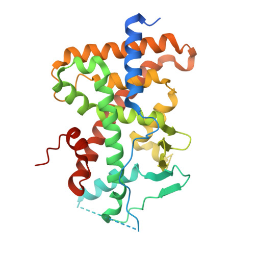 | |
UniProt & NIH Common Fund Data Resources | |||||
PHAROS: O75469 GTEx: ENSG00000144852 | |||||
Entity Groups | |||||
| Sequence Clusters | 30% Identity50% Identity70% Identity90% Identity95% Identity100% Identity | ||||
| UniProt Group | O75469 | ||||
Sequence AnnotationsExpand | |||||
Reference Sequence | |||||
| Ligands 2 Unique | |||||
|---|---|---|---|---|---|
| ID | Chains | Name / Formula / InChI Key | 2D Diagram | 3D Interactions | |
| UK6 (Subject of Investigation/LOI) Download:Ideal Coordinates CCD File | B [auth A] | sclareol C20 H36 O2 XVULBTBTFGYVRC-HHUCQEJWSA-N |  | ||
| GOL Download:Ideal Coordinates CCD File | C [auth A] | GLYCEROL C3 H8 O3 PEDCQBHIVMGVHV-UHFFFAOYSA-N |  | ||
| Length ( Å ) | Angle ( ˚ ) |
|---|---|
| a = 91.828 | α = 90 |
| b = 91.828 | β = 90 |
| c = 85.891 | γ = 90 |
| Software Name | Purpose |
|---|---|
| PHENIX | refinement |
| XDS | data reduction |
| XDS | data scaling |
| MOLREP | phasing |
| Funding Organization | Location | Grant Number |
|---|---|---|
| Other government | France | ANSES 2018/1/020 |


