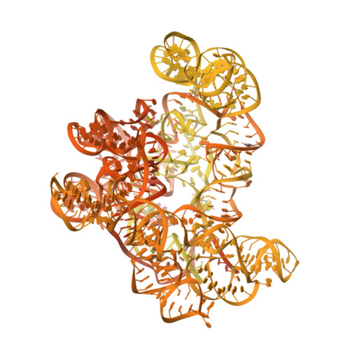
8CF8

Eravacycline bound to the 30S head


Experimental Data Snapshot

  • Method: ELECTRON MICROSCOPY
  • Resolution: 2.20 Å
  • Aggregation State: PARTICLE 
  • Reconstruction Method: SINGLE PARTICLE 

wwPDB Validation   3D Report Full Report


This is version 1.4 of the entry. See complete history


Literature

Structural conservation of antibiotic interaction with ribosomes.

Paternoga, H.Crowe-McAuliffe, C.Bock, L.V.Koller, T.O.Morici, M.Beckert, B.Myasnikov, A.G.Grubmuller, H.Novacek, J.Wilson, D.N.

(2023) Nat Struct Mol Biol 30: 1380-1392

  • DOI: https://doi.org/10.1038/s41594-023-01047-y
  • Primary Citation of Related Structures:  
    8CA7, 8CAI, 8CAM, 8CAZ, 8CEP, 8CEU, 8CF1, 8CF8, 8CGD, 8CGI, 8CGJ, 8CGK, 8CGR, 8CGU, 8CGV

  • PubMed Abstract: 

    The ribosome is a major target for clinically used antibiotics, but multidrug resistant pathogenic bacteria are making our current arsenal of antimicrobials obsolete. Here we present cryo-electron-microscopy structures of 17 distinct compounds from six different antibiotic classes bound to the bacterial ribosome at resolutions ranging from 1.6 to 2.2 Å. The improved resolution enables a precise description of antibiotic-ribosome interactions, encompassing solvent networks that mediate multiple additional interactions between the drugs and their target. Our results reveal a high structural conservation in the binding mode between antibiotics with the same scaffold, including ordered water molecules. Water molecules are visualized within the antibiotic binding sites that are preordered, become ordered in the presence of the drug and that are physically displaced on drug binding. Insight into RNA-ligand interactions will facilitate development of new antimicrobial agents, as well as other RNA-targeting therapies.


  • Organizational Affiliation

    Institute for Biochemistry and Molecular Biology, University of Hamburg, Hamburg, Germany.


Macromolecules

Find similar proteins by:  (by identity cutoff)  |  3D Structure
Entity ID: 2
MoleculeChains Sequence LengthOrganismDetailsImage
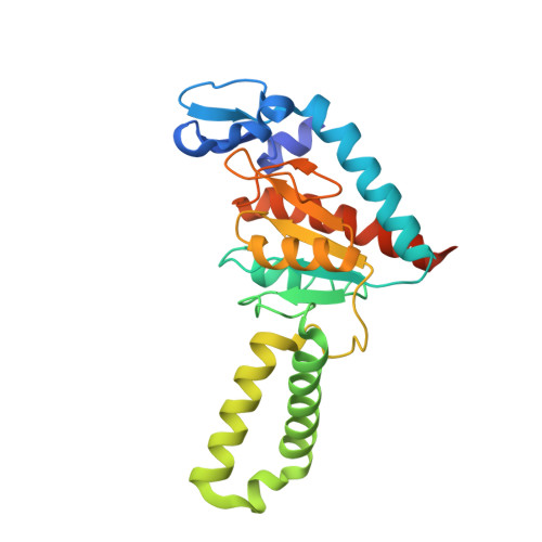
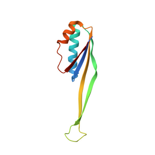
30S ribosomal protein S2241Escherichia coli BW25113Mutation(s): 0 
UniProt
Find proteins for P0A7V0 (Escherichia coli (strain K12))
Explore P0A7V0 
Go to UniProtKB:  P0A7V0
Entity Groups  
Sequence Clusters30% Identity50% Identity70% Identity90% Identity95% Identity100% Identity
UniProt GroupP0A7V0
Sequence Annotations
Expand
  • Reference Sequence
Find similar proteins by:  (by identity cutoff)  |  3D Structure
Entity ID: 3
MoleculeChains Sequence LengthOrganismDetailsImage
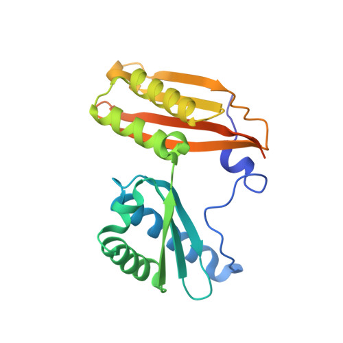
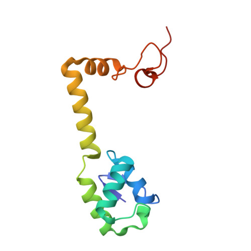
Small ribosomal subunit protein uS3233Escherichia coli BW25113Mutation(s): 0 
UniProt
Find proteins for P0A7V3 (Escherichia coli (strain K12))
Explore P0A7V3 
Go to UniProtKB:  P0A7V3
Entity Groups  
Sequence Clusters30% Identity50% Identity70% Identity90% Identity95% Identity100% Identity
UniProt GroupP0A7V3
Sequence Annotations
Expand
  • Reference Sequence
Find similar proteins by:  (by identity cutoff)  |  3D Structure
Entity ID: 4
MoleculeChains Sequence LengthOrganismDetailsImage
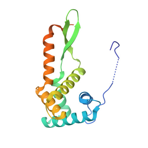
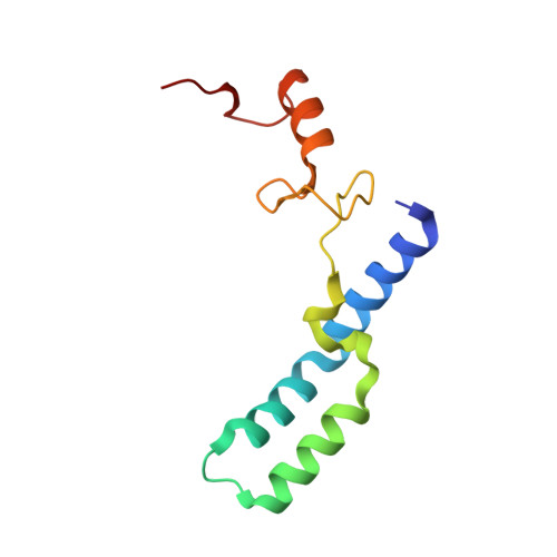
30S ribosomal protein S7D [auth G]179Escherichia coli BW25113Mutation(s): 0 
UniProt
Find proteins for P02359 (Escherichia coli (strain K12))
Explore P02359 
Go to UniProtKB:  P02359
Entity Groups  
Sequence Clusters30% Identity50% Identity70% Identity90% Identity95% Identity100% Identity
UniProt GroupP02359
Sequence Annotations
Expand
  • Reference Sequence
Find similar proteins by:  (by identity cutoff)  |  3D Structure
Entity ID: 5
MoleculeChains Sequence LengthOrganismDetailsImage
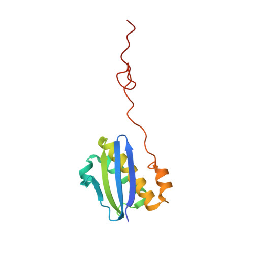
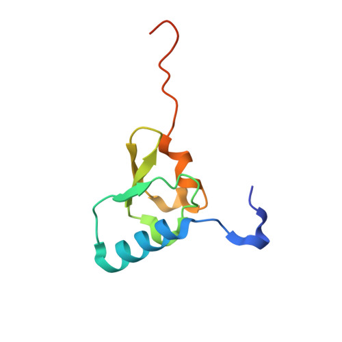
Small ribosomal subunit protein uS9E [auth I]130Escherichia coli BW25113Mutation(s): 0 
UniProt
Find proteins for P0A7X3 (Escherichia coli (strain K12))
Explore P0A7X3 
Go to UniProtKB:  P0A7X3
Entity Groups  
Sequence Clusters30% Identity50% Identity70% Identity90% Identity95% Identity100% Identity
UniProt GroupP0A7X3
Sequence Annotations
Expand
  • Reference Sequence
Find similar proteins by:  (by identity cutoff)  |  3D Structure
Entity ID: 6
MoleculeChains Sequence LengthOrganismDetailsImage
Small ribosomal subunit protein uS10F [auth J]103Escherichia coli BW25113Mutation(s): 0 
UniProt
Find proteins for P0A7R5 (Escherichia coli (strain K12))
Explore P0A7R5 
Go to UniProtKB:  P0A7R5
Entity Groups  
Sequence Clusters30% Identity50% Identity70% Identity90% Identity95% Identity100% Identity
UniProt GroupP0A7R5
Sequence Annotations
Expand
  • Reference Sequence
Find similar proteins by:  (by identity cutoff)  |  3D Structure
Entity ID: 7
MoleculeChains Sequence LengthOrganismDetailsImage
Small ribosomal subunit protein uS13G [auth M]118Escherichia coli BW25113Mutation(s): 0 
UniProt
Find proteins for P0A7S9 (Escherichia coli (strain K12))
Explore P0A7S9 
Go to UniProtKB:  P0A7S9
Entity Groups  
Sequence Clusters30% Identity50% Identity70% Identity90% Identity95% Identity100% Identity
UniProt GroupP0A7S9
Sequence Annotations
Expand
  • Reference Sequence
Find similar proteins by:  (by identity cutoff)  |  3D Structure
Entity ID: 8
MoleculeChains Sequence LengthOrganismDetailsImage
Small ribosomal subunit protein uS14H [auth N]101Escherichia coli BW25113Mutation(s): 0 
UniProt
Find proteins for P0AG59 (Escherichia coli (strain K12))
Explore P0AG59 
Go to UniProtKB:  P0AG59
Entity Groups  
Sequence Clusters30% Identity50% Identity70% Identity90% Identity95% Identity100% Identity
UniProt GroupP0AG59
Sequence Annotations
Expand
  • Reference Sequence
Find similar proteins by:  (by identity cutoff)  |  3D Structure
Entity ID: 9
MoleculeChains Sequence LengthOrganismDetailsImage
Small ribosomal subunit protein uS19I [auth S]92Escherichia coli BW25113Mutation(s): 0 
UniProt
Find proteins for P0A7U3 (Escherichia coli (strain K12))
Explore P0A7U3 
Go to UniProtKB:  P0A7U3
Entity Groups  
Sequence Clusters30% Identity50% Identity70% Identity90% Identity95% Identity100% Identity
UniProt GroupP0A7U3
Sequence Annotations
Expand
  • Reference Sequence
Find similar nucleic acids by:  (by identity cutoff)  |  3D Structure
Entity ID: 1
MoleculeChains LengthOrganismImage
16S rRNA1,540Escherichia coli BW25113
Sequence Annotations
Expand
  • Reference Sequence
Small Molecules
Ligands 3 Unique
IDChains Name / Formula / InChI Key2D Diagram3D Interactions
YQM (Subject of Investigation/LOI)
Query on YQM

Download Ideal Coordinates CCD File 
J [auth A]Eravacycline
C27 H31 F N4 O8
AKLMFDDQCHURPW-ISIOAQNYSA-N
K
Query on K

Download Ideal Coordinates CCD File 
AB [auth A]
BB [auth M]
QA [auth A]
RA [auth A]
SA [auth A]
AB [auth A],
BB [auth M],
QA [auth A],
RA [auth A],
SA [auth A],
TA [auth A],
UA [auth A],
VA [auth A],
WA [auth A],
XA [auth A],
YA [auth A],
ZA [auth A]
POTASSIUM ION
K
NPYPAHLBTDXSSS-UHFFFAOYSA-N
MG
Query on MG

Download Ideal Coordinates CCD File 
AA [auth A]
BA [auth A]
CA [auth A]
DA [auth A]
EA [auth A]
AA [auth A],
BA [auth A],
CA [auth A],
DA [auth A],
EA [auth A],
FA [auth A],
GA [auth A],
HA [auth A],
IA [auth A],
JA [auth A],
K [auth A],
KA [auth A],
L [auth A],
LA [auth A],
M [auth A],
MA [auth A],
N [auth A],
NA [auth A],
O [auth A],
OA [auth A],
P [auth A],
PA [auth A],
Q [auth A],
R [auth A],
S [auth A],
T [auth A],
U [auth A],
V [auth A],
W [auth A],
X [auth A],
Y [auth A],
Z [auth A]
MAGNESIUM ION
Mg
JLVVSXFLKOJNIY-UHFFFAOYSA-N
Experimental Data & Validation

Experimental Data

  • Method: ELECTRON MICROSCOPY
  • Resolution: 2.20 Å
  • Aggregation State: PARTICLE 
  • Reconstruction Method: SINGLE PARTICLE 
EM Software:
TaskSoftware PackageVersion
MODEL REFINEMENTREFMAC5.8.0415

Structure Validation

View Full Validation Report


Entry History & Funding Information

Deposition Data


Funding OrganizationLocationGrant Number
Other governmentDLR01Kl1820
iNEXT-DiscoveryEuropean Union871037
Other governmentLM2018127

Revision History  (Full details and data files)

  • Version 1.0: 2023-08-02
    Type: Initial release
  • Version 1.1: 2023-08-09
    Changes: Database references
  • Version 1.2: 2023-08-16
    Changes: Data collection, Database references
  • Version 1.3: 2023-09-20
    Changes: Database references
  • Version 1.4: 2024-04-24
    Changes: Data collection