8CAV

Discovery of the lanthipeptide Curvocidin and structural insights into its trifunctional synthetase CuvL


Experimental Data Snapshot

  • Method: X-RAY DIFFRACTION
  • Resolution: 2.87 Å
  • R-Value Free: 0.293 
  • R-Value Work: 0.237 
  • R-Value Observed: 0.240 

Starting Model: experimental
View more details

wwPDB Validation   3D Report Full Report


Ligand Structure Quality Assessment 


This is version 1.1 of the entry. See complete history


Literature

Discovery of the Lanthipeptide Curvocidin and Structural Insights into its Trifunctional Synthetase CuvL.

Sigurdsson, A.Martins, B.M.Duttmann, S.A.Jasyk, M.Dimos-Rohl, B.Schopf, F.Gemander, M.Knittel, C.H.Schnegotzki, R.Schmid, B.Kosol, S.Pommerening, L.Gonzales-Viegaz, M.Seidel, M.Hugelland, M.Leimkuhler, S.Dobbek, H.Mainz, A.Sussmuth, R.D.

(2023) Angew Chem Int Ed Engl 62: e202302490-e202302490

  • DOI: https://doi.org/10.1002/anie.202302490
  • Primary Citation of Related Structures:  
    8CAR, 8CAV

  • PubMed Abstract: 

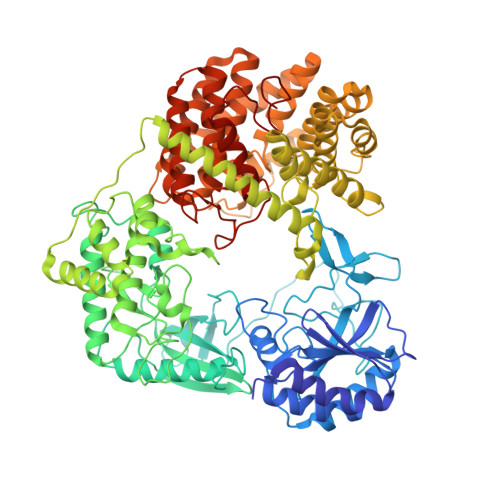
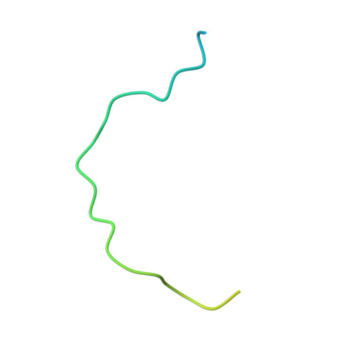
    Lanthipeptides are ribosomally-synthesized natural products from bacteria featuring stable thioether-crosslinks and various bioactivities. Herein, we report on a new clade of tricyclic class-IV lanthipeptides with curvocidin from Thermomonospora curvata as its first representative. We obtained crystal structures of the corresponding lanthipeptide synthetase CuvL that showed a circular arrangement of its kinase, lyase and cyclase domains, forming a central reaction chamber for the iterative substrate processing involving nine catalytic steps. The combination of experimental data and artificial intelligence-based structural models identified the N-terminal subdomain of the kinase domain as the primary site of substrate recruitment. The ribosomal precursor peptide of curvocidin employs an amphipathic α-helix in its leader region as an anchor to CuvL, while its substrate core shuttles within the central reaction chamber. Our study thus reveals general principles of domain organization and substrate recruitment of class-IV and class-III lanthipeptide synthetases.


  • Organizational Affiliation

    Fakultät II-Institut für Chemie, Technische Universität Berlin, Straße des 17. Juni 124, 10623, Berlin, Germany.


Macromolecules
Find similar proteins by:  (by identity cutoff)  |  3D Structure
Entity ID: 1
MoleculeChains Sequence LengthOrganismDetailsImage
Serine/threonine protein kinase
A, B
865Thermomonospora curvataMutation(s): 0 
Gene Names: Tcur_0376
EC: 2.7.11.1
UniProt
Find proteins for D1A2F7 (Thermomonospora curvata (strain ATCC 19995 / DSM 43183 / JCM 3096 / KCTC 9072 / NBRC 15933 / NCIMB 10081 / Henssen B9))
Explore D1A2F7 
Go to UniProtKB:  D1A2F7
Entity Groups  
Sequence Clusters30% Identity50% Identity70% Identity90% Identity95% Identity100% Identity
UniProt GroupD1A2F7
Sequence Annotations
Expand
  • Reference Sequence
Find similar proteins by:  (by identity cutoff)  |  3D Structure
Entity ID: 2
MoleculeChains Sequence LengthOrganismDetailsImage
CuvAC [auth D],
D [auth G]
51Thermomonospora curvataMutation(s): 0 
Gene Names: Tcur_0378
UniProt
Find proteins for D1A2F9 (Thermomonospora curvata (strain ATCC 19995 / DSM 43183 / JCM 3096 / KCTC 9072 / NBRC 15933 / NCIMB 10081 / Henssen B9))
Explore D1A2F9 
Go to UniProtKB:  D1A2F9
Entity Groups  
Sequence Clusters30% Identity50% Identity70% Identity90% Identity95% Identity100% Identity
UniProt GroupD1A2F9
Sequence Annotations
Expand
  • Reference Sequence
Small Molecules
Ligands 3 Unique
IDChains Name / Formula / InChI Key2D Diagram3D Interactions
ANP (Subject of Investigation/LOI)
Query on ANP

Download Ideal Coordinates CCD File 
G [auth A],
J [auth B]
PHOSPHOAMINOPHOSPHONIC ACID-ADENYLATE ESTER
C10 H17 N6 O12 P3
PVKSNHVPLWYQGJ-KQYNXXCUSA-N
MPD
Query on MPD

Download Ideal Coordinates CCD File 
E [auth A],
F [auth A],
I [auth B],
K [auth B]
(4S)-2-METHYL-2,4-PENTANEDIOL
C6 H14 O2
SVTBMSDMJJWYQN-YFKPBYRVSA-N
MG
Query on MG

Download Ideal Coordinates CCD File 
H [auth A],
L [auth B]
MAGNESIUM ION
Mg
JLVVSXFLKOJNIY-UHFFFAOYSA-N
Experimental Data & Validation

Experimental Data

  • Method: X-RAY DIFFRACTION
  • Resolution: 2.87 Å
  • R-Value Free: 0.293 
  • R-Value Work: 0.237 
  • R-Value Observed: 0.240 
  • Space Group: P 1 21 1
Unit Cell:
Length ( Å )Angle ( ˚ )
a = 84.37α = 90
b = 116.13β = 106.25
c = 97.41γ = 90
Software Package:
Software NamePurpose
autoXDSdata collection
PHENIXphasing
PHENIXrefinement
Cootmodel building

Structure Validation

View Full Validation Report


Ligand Structure Quality Assessment 


Entry History & Funding Information

Deposition Data


Funding OrganizationLocationGrant Number
German Research Foundation (DFG)GermanyEXC 2008 390540038

Revision History  (Full details and data files)

  • Version 1.0: 2023-06-14
    Type: Initial release
  • Version 1.1: 2024-10-23
    Changes: Data collection, Structure summary