8C7H | pdb_00008c7h

Cryo-EM Map of the latTGF-beta LHG-10 Fab complex


Experimental Data Snapshot

  • Method: ELECTRON MICROSCOPY
  • Resolution: 2.70 Å
  • Aggregation State: PARTICLE 
  • Reconstruction Method: SINGLE PARTICLE 

wwPDB Validation 3D Report Full Report

Validation slider image for 8C7H

This is version 1.4 of the entry. See complete history

Literature

Anti-GARP Antibodies Inhibit Release of TGF-beta by Regulatory T Cells via Different Modes of Action, but Do Not Influence Their Function In Vitro.

Igney, F.H.Ebenhoch, R.Schiele, F.Nar, H.

(2023) Immunohorizons 7: 200-212

  • DOI: https://doi.org/10.4049/immunohorizons.2200072
  • Primary Citation Related Structures: 
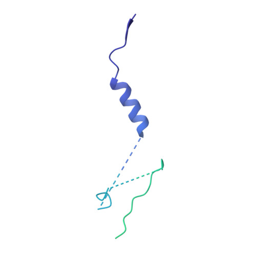
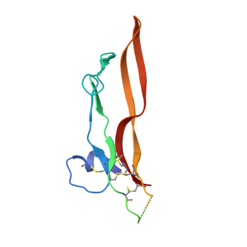
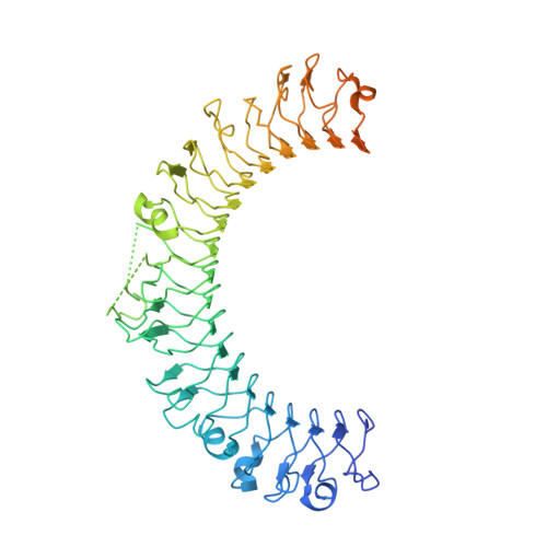
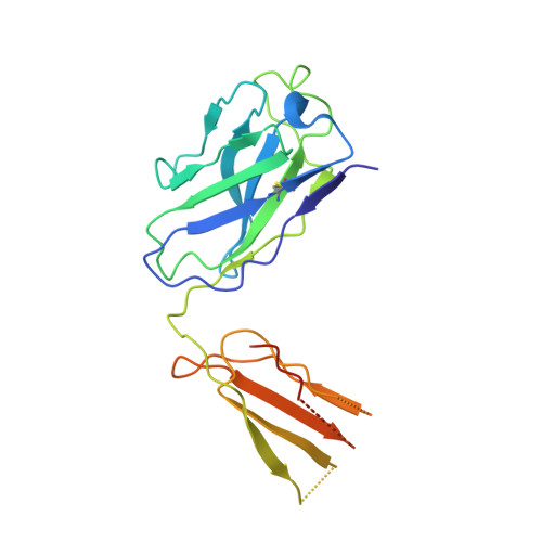
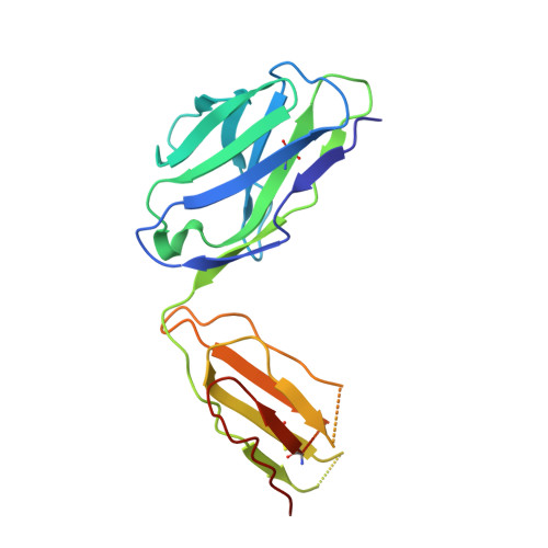
    8C7H

  • PubMed Abstract: 

    Regulatory T cells (Treg) play a critical role in controlling immune responses in diseases such as cancer or autoimmunity. Activated Treg express the membrane protein GARP (LRRC32) in complex with the latent form of the immunosuppressive cytokine TGF-β (L-TGF-β). In this study, we confirmed that active TGF-β was generated from its latent form in an integrin-dependent manner and induced TGF-β receptor signaling in activated human Treg. We studied a series of Abs targeting the L-TGF-β/GARP complex with distinct binding modes. We found that TGF-β receptor signaling could be inhibited by anti-TGF-β and by some, but not all, Abs against the L-TGF-β/GARP complex. Cryogenic electron microscopy structures of three L-TGF-β/GARP complex-targeting Abs revealed their distinct epitopes and allowed us to elucidate how they achieve blockade of TGF-β activation. Three different modes of action were identified, including a novel unusual mechanism of a GARP-binding Ab. However, blockade of GARP or TGF-β by Abs did not influence the suppressive activity of human Treg in vitro. We were also not able to confirm a prominent role of GARP in other functions of human Treg, such as FOXP3 induction and Treg stability. These data show that the GARP/TGF-β axis can be targeted pharmacologically in different ways, but further studies are necessary to understand its complexity and to unleash its therapeutic potential.


  • Organizational Affiliation
    • Discovery Research, Cancer Immunology & Immune Modulation, Boehringer Ingelheim Pharma GmbH & Co. KG, Biberach an der Riss, Germany.

Macromolecule Content 

  • Total Structure Weight: 197.42 kDa 
  • Atom Count: 9,172 
  • Modeled Residue Count: 1,208 
  • Deposited Residue Count: 1,764 
  • Unique protein chains: 5

Macromolecules

Find similar proteins by:|  3D Structure
Entity ID: 1
MoleculeChains  Sequence LengthOrganismDetailsImage
Transforming growth factor beta-1
A, C
249Homo sapiensMutation(s): 0 
Gene Names: TGFB1TGFB
UniProt & NIH Common Fund Data Resources
Find proteins for P01137 (Homo sapiens)
Explore P01137 
Go to UniProtKB:  P01137
PHAROS:  P01137
GTEx:  ENSG00000105329 
Entity Groups
Sequence Clusters30% Identity50% Identity70% Identity90% Identity95% Identity100% Identity
UniProt GroupP01137
Sequence Annotations
Expand
Reference Sequence
Find similar proteins by:|  3D Structure
Entity ID: 2
MoleculeChains  Sequence LengthOrganismDetailsImage
Transforming growth factor beta-1
B, D
112Homo sapiensMutation(s): 0 
Gene Names: TGFB1TGFB
UniProt & NIH Common Fund Data Resources
Find proteins for P01137 (Homo sapiens)
Explore P01137 
Go to UniProtKB:  P01137
PHAROS:  P01137
GTEx:  ENSG00000105329 
Entity Groups
Sequence Clusters30% Identity50% Identity70% Identity90% Identity95% Identity100% Identity
UniProt GroupP01137
Sequence Annotations
Expand
Reference Sequence
Find similar proteins by:|  3D Structure
Entity ID: 3
MoleculeChains  Sequence LengthOrganismDetailsImage
Transforming growth factor beta activator LRRC32E [auth I]601Homo sapiensMutation(s): 0 
Gene Names: LRRC32D11S833E
UniProt & NIH Common Fund Data Resources
Find proteins for Q14392 (Homo sapiens)
Explore Q14392 
Go to UniProtKB:  Q14392
PHAROS:  Q14392
GTEx:  ENSG00000137507 
Entity Groups
Sequence Clusters30% Identity50% Identity70% Identity90% Identity95% Identity100% Identity
UniProt GroupQ14392
Glycosylation
Glycosylation Sites: 3Go to GlyGen: Q14392-1
Sequence Annotations
Expand
Reference Sequence
Find similar proteins by:|  3D Structure
Entity ID: 4
MoleculeChains  Sequence LengthOrganismDetailsImage
28G11 Fab heavy chainF [auth L]227Homo sapiensMutation(s): 0 
Find similar proteins by:|  3D Structure
Entity ID: 5
MoleculeChains  Sequence LengthOrganismDetailsImage
28G11 Fab light chainG [auth K]214Homo sapiensMutation(s): 0 

Oligosaccharides

Help  
Entity ID: 6
MoleculeChains Length2D Diagram GlycosylationD Interactions
beta-D-mannopyranose-(1-4)-2-acetamido-2-deoxy-beta-D-glucopyranose-(1-4)-2-acetamido-2-deoxy-beta-D-glucopyranoseH [auth E],
I [auth F],
J [auth G]
3N-Glycosylation
Glycosylation Resources
GlyTouCan: G15407YE
GlyCosmos: G15407YE
GlyGen: G15407YE

Experimental Data & Validation

Experimental Data

  • Method: ELECTRON MICROSCOPY
  • Resolution: 2.70 Å
  • Aggregation State: PARTICLE 
  • Reconstruction Method: SINGLE PARTICLE 
EM Software:
TaskSoftware PackageVersion
MODEL REFINEMENTPHENIX

Structure Validation

View Full Validation Report


Entry History 

& Funding Information

Deposition Data


Funding OrganizationLocationGrant Number
Not funded--

Revision History  (Full details and data files)

  • Version 1.0: 2023-03-01
    Type: Initial release
  • Version 1.1: 2023-03-29
    Changes: Database references
  • Version 1.2: 2024-10-09
    Changes: Data collection, Structure summary
  • Version 1.3: 2024-11-06
    Changes: Data collection, Structure summary
  • Version 1.4: 2025-07-09
    Changes: Data collection