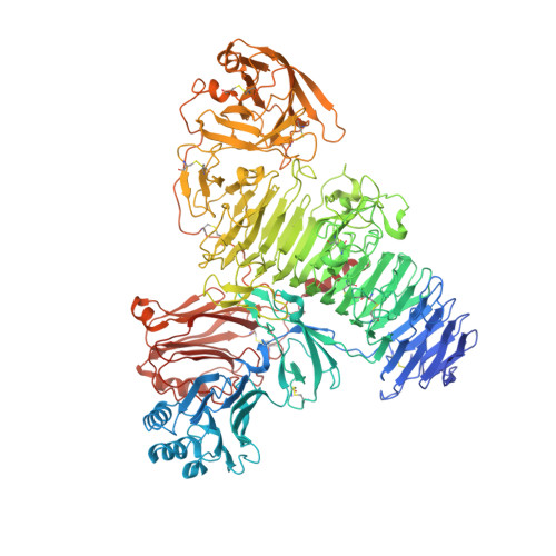
8C6I

TMEM2 ectodomain


Experimental Data Snapshot

  • Method: X-RAY DIFFRACTION
  • Resolution: 3.50 Å
  • R-Value Free: 0.319 
  • R-Value Work: 0.265 
  • R-Value Observed: 0.268 

wwPDB Validation   3D Report Full Report


Ligand Structure Quality Assessment 


This is version 1.1 of the entry. See complete history


Literature

Structure of the transmembrane protein 2 (TMEM2) ectodomain and its apparent lack of hyaluronidase activity.

Niu, M.McGrath, M.Sammon, D.Gardner, S.Morgan, R.M.Di Maio, A.Liu, Y.Bubeck, D.Hohenester, E.

(2023) Wellcome Open Res 8: 76-76

  • DOI: https://doi.org/10.12688/wellcomeopenres.18937.2
  • Primary Citation of Related Structures:  
    8C6I

  • PubMed Abstract: 

    Background: Hyaluronic acid (HA) is a major polysaccharide component of the extracellular matrix. HA has essential functions in tissue architecture and the regulation of cell behaviour. HA turnover needs to be finely balanced. Increased HA degradation is associated with cancer, inflammation, and other pathological situations. Transmembrane protein 2 (TMEM2) is a cell surface protein that has been reported to degrade HA into ~5 kDa fragments and play an essential role in systemic HA turnover. Methods: We produced the soluble TMEM2 ectodomain (residues 106-1383; sTMEM2) in human embryonic kidney cells (HEK293) and determined its structure using X-ray crystallography. We tested sTMEM2 hyaluronidase activity using fluorescently labelled HA and size fractionation of reaction products. We tested HA binding in solution and using a glycan microarray. Results: Our crystal structure of sTMEM2 confirms a remarkably accurate prediction by AlphaFold. sTMEM2 contains a parallel β-helix typical of other polysaccharide-degrading enzymes, but an active site cannot be assigned with confidence. A lectin-like domain is inserted into the β-helix and predicted to be functional in carbohydrate binding. A second lectin-like domain at the C-terminus is unlikely to bind carbohydrates. We did not observe HA binding in two assay formats, suggesting a modest affinity at best. Unexpectedly, we were unable to observe any HA degradation by sTMEM2. Our negative results set an upper limit for k cat of approximately 10 -5 min -1 . Conclusions: Although sTMEM2 contains domain types consistent with its suggested role in TMEM2 degradation, its hyaluronidase activity was undetectable. HA degradation by TMEM2 may require additional proteins and/or localisation at the cell surface.


  • Organizational Affiliation

    Department of Life Sciences, Imperial College London, London, SW7 2AZ, UK.


Macromolecules
Find similar proteins by:  (by identity cutoff)  |  3D Structure
Entity ID: 1
MoleculeChains Sequence LengthOrganismDetailsImage
Cell surface hyaluronidase1,301Homo sapiensMutation(s): 0 
Gene Names: CEMIP2KIAA1412TMEM2
EC: 3.2.1.35
UniProt & NIH Common Fund Data Resources
Find proteins for Q9UHN6 (Homo sapiens)
Explore Q9UHN6 
Go to UniProtKB:  Q9UHN6
PHAROS:  Q9UHN6
GTEx:  ENSG00000135048 
Entity Groups  
Sequence Clusters30% Identity50% Identity70% Identity90% Identity95% Identity100% Identity
UniProt GroupQ9UHN6
Sequence Annotations
Expand
  • Reference Sequence
Oligosaccharides

Help

Entity ID: 2
MoleculeChains Length2D Diagram Glycosylation3D Interactions
alpha-D-mannopyranose-(1-6)-beta-D-mannopyranose-(1-4)-2-acetamido-2-deoxy-beta-D-glucopyranose-(1-4)-2-acetamido-2-deoxy-beta-D-glucopyranose
B, D
4N-Glycosylation
Glycosylation Resources
GlyTouCan:  G22573RC
GlyCosmos:  G22573RC
GlyGen:  G22573RC
Entity ID: 3
MoleculeChains Length2D Diagram Glycosylation3D Interactions
2-acetamido-2-deoxy-beta-D-glucopyranose-(1-4)-2-acetamido-2-deoxy-beta-D-glucopyranose
C, H
2N-Glycosylation
Glycosylation Resources
GlyTouCan:  G42666HT
GlyCosmos:  G42666HT
GlyGen:  G42666HT
Entity ID: 4
MoleculeChains Length2D Diagram Glycosylation3D Interactions
beta-D-mannopyranose-(1-4)-2-acetamido-2-deoxy-beta-D-glucopyranose-(1-4)-2-acetamido-2-deoxy-beta-D-glucopyranose
E
3N-Glycosylation
Glycosylation Resources
GlyTouCan:  G15407YE
GlyCosmos:  G15407YE
GlyGen:  G15407YE
Entity ID: 5
MoleculeChains Length2D Diagram Glycosylation3D Interactions
alpha-D-mannopyranose-(1-6)-beta-D-mannopyranose-(1-4)-2-acetamido-2-deoxy-beta-D-glucopyranose-(1-4)-[alpha-L-fucopyranose-(1-6)]2-acetamido-2-deoxy-beta-D-glucopyranose
F
5N-Glycosylation
Glycosylation Resources
GlyTouCan:  G00395TQ
GlyCosmos:  G00395TQ
GlyGen:  G00395TQ
Entity ID: 6
MoleculeChains Length2D Diagram Glycosylation3D Interactions
alpha-D-mannopyranose-(1-3)-beta-D-mannopyranose-(1-4)-2-acetamido-2-deoxy-beta-D-glucopyranose-(1-4)-2-acetamido-2-deoxy-beta-D-glucopyranose
G
4N-Glycosylation
Glycosylation Resources
GlyTouCan:  G81315DD
GlyCosmos:  G81315DD
GlyGen:  G81315DD
Experimental Data & Validation

Experimental Data

  • Method: X-RAY DIFFRACTION
  • Resolution: 3.50 Å
  • R-Value Free: 0.319 
  • R-Value Work: 0.265 
  • R-Value Observed: 0.268 
  • Space Group: P 42 21 2
Unit Cell:
Length ( Å )Angle ( ˚ )
a = 184.76α = 90
b = 184.76β = 90
c = 105.398γ = 90
Software Package:
Software NamePurpose
PHENIXrefinement
DIALSdata reduction
DIALSdata scaling
PHENIXphasing

Structure Validation

View Full Validation Report


Ligand Structure Quality Assessment 


Entry History & Funding Information

Deposition Data


Funding OrganizationLocationGrant Number
Wellcome TrustUnited Kingdom101748/Z/13/Z

Revision History  (Full details and data files)

  • Version 1.0: 2023-02-01
    Type: Initial release
  • Version 1.1: 2023-06-07
    Changes: Database references