







 Download Mendeley
Dark state of PAS-GAF fragment from Deinococcus radiodurans phytochrome
Madan Kumar, S., Sebastian, W.To be published.
Experimental Data Snapshot
Starting Model: experimental
View more details
Entity ID: 1 | |||||
|---|---|---|---|---|---|
| Molecule | Chains | Sequence Length | Organism | Details | Image |
| Bacteriophytochrome | A [auth B], B [auth A] | 343 | Deinococcus radiodurans | Mutation(s): 0 Gene Names: bphP, DR_A0050 EC: 2.7.13.3 |  |
UniProt | |||||
Entity Groups | |||||
| Sequence Clusters | 30% Identity50% Identity70% Identity90% Identity95% Identity100% Identity | ||||
| UniProt Group | Q9RZA4 | ||||
Sequence AnnotationsExpand | |||||
Reference Sequence | |||||
| Ligands 1 Unique | |||||
|---|---|---|---|---|---|
| ID | Chains | Name / Formula / InChI Key | 2D Diagram | 3D Interactions | |
| LBV (Subject of Investigation/LOI) Download:Ideal Coordinates CCD File | C [auth B], D [auth A] | 3-[2-[(Z)-[3-(2-carboxyethyl)-5-[(Z)-(4-ethenyl-3-methyl-5-oxidanylidene-pyrrol-2-ylidene)methyl]-4-methyl-pyrrol-1-ium
-2-ylidene]methyl]-5-[(Z)-[(3E)-3-ethylidene-4-methyl-5-oxidanylidene-pyrrolidin-2-ylidene]methyl]-4-methyl-1H-pyrrol-3-
yl]propanoic acid C33 H37 N4 O6 DKMLMZVDTGOEGU-ISEYCTJISA-O | 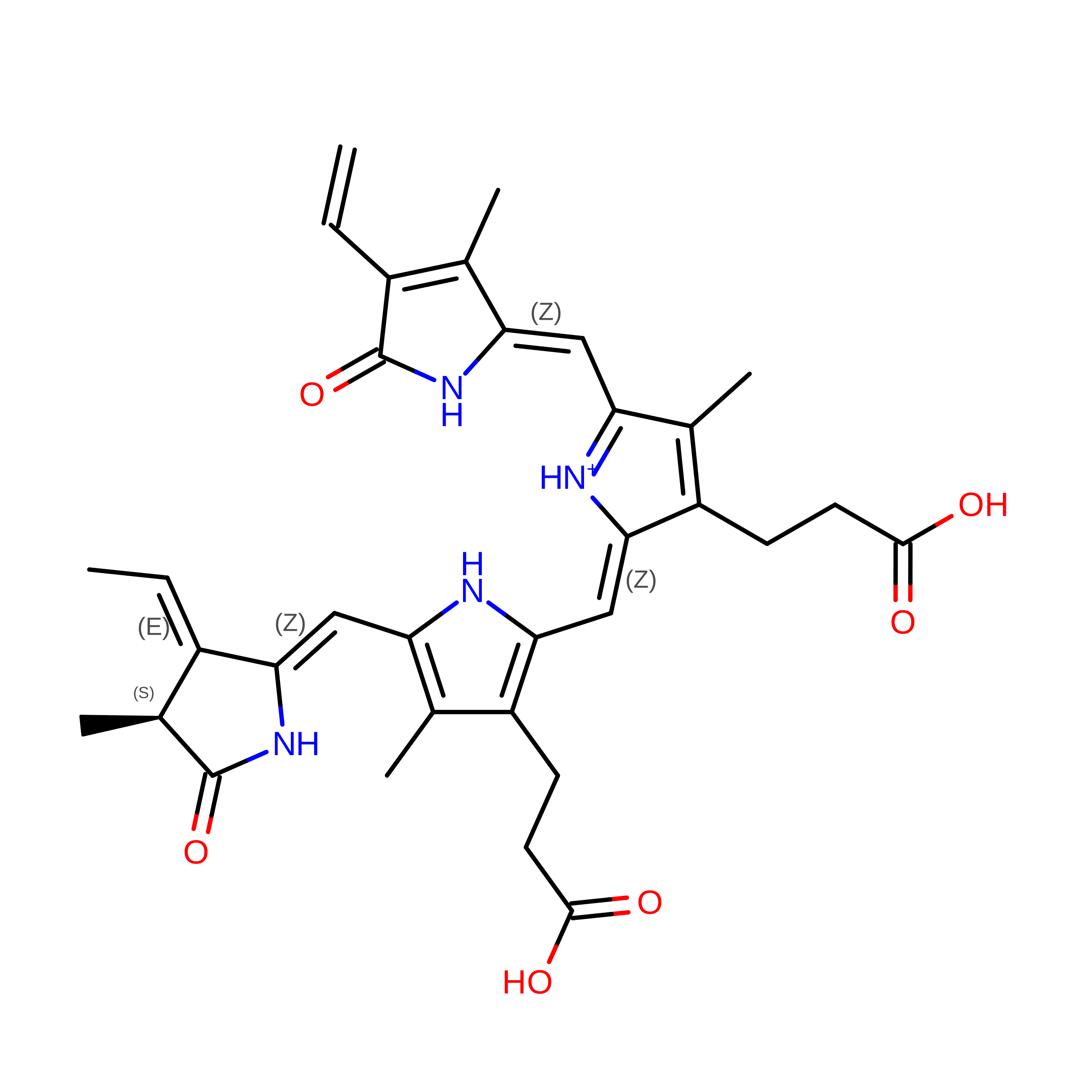 | ||
| Length ( Å ) | Angle ( ˚ ) |
|---|---|
| a = 54.95 | α = 90 |
| b = 116.5 | β = 90 |
| c = 117.7 | γ = 90 |
| Software Name | Purpose |
|---|---|
| PHENIX | refinement |
| CrystFEL | data reduction |
| CrystFEL | data scaling |
| PHASER | phasing |
| Funding Organization | Location | Grant Number |
|---|---|---|
| European Research Council (ERC) | European Union | 279944 |
| Japan Agency for Medical Research and Development (AMED) | Japan | -- |
| Academy of Finland | Finland | 285461 |








