Computational structure-based approach to study chimeric antigens using a new protein scaffold displaying foreign epitopes.
Cappelli, L., Cinelli, P., Perrotta, A., Veggi, D., Audagnotto, M., Tuscano, G., Pansegrau, W., Bartolini, E., Rinaudo, D., Cozzi, R.(2024) FASEB J 38: e23326-e23326
- PubMed: 38019196 Search on PubMed
- DOI: https://doi.org/10.1096/fj.202202130R
- Primary Citation Related Structures:
8C27 - PubMed Abstract:
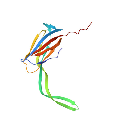
The identification and recombinant production of functional antigens and/or epitopes of pathogens represent a crucial step for the development of an effective protein-based vaccine. Many vaccine targets are outer membrane proteins anchored into the lipidic bilayer through an extended hydrophobic portion making their recombinant production challenging. Moreover, only the extracellular loops, and not the hydrophobic regions, are naturally exposed to the immune system. In this work, the Domain 3 (D3) from Group B Streptococcus (GBS) pilus 2a backbone protein has been identified and engineered to be used as a scaffold for the display of extracellular loops of two Neisseria gonorrhoeae membrane proteins (PorB.1b and OpaB). A computational structure-based approach has been applied to the design of both the scaffold and the model antigens. Once identified the best D3 engineerable site, several different chimeric D3 displaying PorB.1b and OpaB extracellular loops were produced as soluble proteins. Each molecule has been characterized in terms of solubility, stability, and ability to correctly display the foreign epitope. This antigen dissection strategy allowed the identification of most immunogenic extracellular loops of both PorB.1b and OpaB gonococcal antigens. The crystal structure of chimeric D3 displaying PorB.1b immunodominant loop has been obtained confirming that the engineerization did not alter the predicted native structure of this epitope. Taken together, the reported data suggest that D3 is a novel protein scaffold for epitope insertion and display, and a valid alternative to the production of whole membrane protein antigens. Finally, this work describes a generalized computational structure-based approach for the identification, design, and dissection of epitopes in target antigens through chimeric proteins.
- Dipartimento di Farmacia e Biotecnologie - FaBiT, University of Bologna, Bologna, Italy.
Organizational Affiliation: