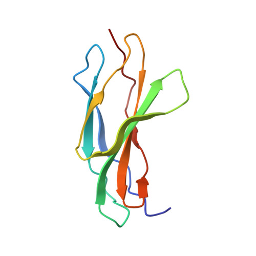
8BW6

Titin FnIII-domain I110 (I/A6) from the MIR region


Experimental Data Snapshot

  • Method: X-RAY DIFFRACTION
  • Resolution: 1.95 Å
  • R-Value Free: 0.254 
  • R-Value Work: 0.217 
  • R-Value Observed: 0.219 

Starting Model: experimental
View more details

wwPDB Validation   3D Report Full Report


This is version 1.2 of the entry. See complete history


Literature

Immunological and Structural Characterization of Titin Main Immunogenic Region; I110 Domain Is the Target of Titin Antibodies in Myasthenia Gravis.

Stergiou, C.Williams, R.Fleming, J.R.Zouvelou, V.Ninou, E.Andreetta, F.Rinaldi, E.Simoncini, O.Mantegazza, R.Bogomolovas, J.Tzartos, J.Labeit, S.Mayans, O.Tzartos, S.

(2023) Biomedicines 11

  • DOI: https://doi.org/10.3390/biomedicines11020449
  • Primary Citation of Related Structures:  
    8BVO, 8BW6, 8BXR

  • PubMed Abstract: 

    Myasthenia gravis (MG) is an autoimmune disease caused by antibodies targeting the neuromuscular junction (NJ) of skeletal muscles. The major MG autoantigen is nicotinic acetylcholine receptor. Other autoantigens at the NJ include MuSK, LRP4 and agrin. Autoantibodies to the intra-sarcomeric striated muscle-specific gigantic protein titin, although not directed to the NJ, are invaluable biomarkers for thymoma and MG disease severity. Thymus and thymoma are critical in MG mechanisms and management. Titin autoantibodies bind to a 30 KDa titin segment, the main immunogenic region (MIR), consisting of an Ig-FnIII-FnIII 3-domain tandem, termed I109-I111. In this work, we further resolved the localization of titin epitope(s) to facilitate the development of more specific anti-titin diagnostics. For this, we expressed protein samples corresponding to 8 MIR and non-MIR titin fragments and tested 77 anti-titin sera for antibody binding using ELISA, competition experiments and Western blots. All anti-MIR antibodies were bound exclusively to the central MIR domain, I110, and to its containing titin segments. Most antibodies were bound also to SDS-denatured I110 on Western blots, suggesting that their epitope(s) are non-conformational. No significant difference was observed between thymoma and non-thymoma patients or between early- and late-onset MG. In addition, atomic 3D-structures of the MIR and its subcomponents were elucidated using X-ray crystallography. These immunological and structural data will allow further studies into the atomic determinants underlying titin-based autoimmunity, improved diagnostics and how to eventually treat titin autoimmunity associated co-morbidities.


  • Organizational Affiliation

    Tzartos NeuroDiagnostics, 115 23 Athens, Greece.


Macromolecules
Find similar proteins by:  (by identity cutoff)  |  3D Structure
Entity ID: 1
MoleculeChains Sequence LengthOrganismDetailsImage
Titin99Homo sapiensMutation(s): 0 
Gene Names: TTN
EC: 2.7.11.1
UniProt & NIH Common Fund Data Resources
Find proteins for Q8WZ42 (Homo sapiens)
Explore Q8WZ42 
Go to UniProtKB:  Q8WZ42
PHAROS:  Q8WZ42
GTEx:  ENSG00000155657 
Entity Groups  
Sequence Clusters30% Identity50% Identity70% Identity90% Identity95% Identity100% Identity
UniProt GroupQ8WZ42
Sequence Annotations
Expand
  • Reference Sequence
Small Molecules
Ligands 3 Unique
IDChains Name / Formula / InChI Key2D Diagram3D Interactions
PEG
Query on PEG

Download Ideal Coordinates CCD File 
E [auth A]DI(HYDROXYETHYL)ETHER
C4 H10 O3
MTHSVFCYNBDYFN-UHFFFAOYSA-N
EDO
Query on EDO

Download Ideal Coordinates CCD File 
B [auth A]
C [auth A]
D [auth A]
G [auth A]
H [auth A]
B [auth A],
C [auth A],
D [auth A],
G [auth A],
H [auth A],
I [auth A],
J [auth A]
1,2-ETHANEDIOL
C2 H6 O2
LYCAIKOWRPUZTN-UHFFFAOYSA-N
CA
Query on CA

Download Ideal Coordinates CCD File 
F [auth A]CALCIUM ION
Ca
BHPQYMZQTOCNFJ-UHFFFAOYSA-N
Experimental Data & Validation

Experimental Data

  • Method: X-RAY DIFFRACTION
  • Resolution: 1.95 Å
  • R-Value Free: 0.254 
  • R-Value Work: 0.217 
  • R-Value Observed: 0.219 
  • Space Group: P 41 21 2
Unit Cell:
Length ( Å )Angle ( ˚ )
a = 45.86α = 90
b = 45.86β = 90
c = 112.02γ = 90
Software Package:
Software NamePurpose
PHENIXrefinement
XDSdata reduction
XSCALEdata scaling
PHASERphasing

Structure Validation

View Full Validation Report


Entry History & Funding Information

Deposition Data


Funding OrganizationLocationGrant Number
British Heart FoundationUnited KingdomPG/13/21/3007
Leducq FoundationFranceTNE-13CVD04

Revision History  (Full details and data files)

  • Version 1.0: 2023-02-15
    Type: Initial release
  • Version 1.1: 2023-03-08
    Changes: Database references
  • Version 1.2: 2024-10-16
    Changes: Data collection, Structure summary