8BTO | pdb_00008bto

Helical structure of BcThsA in complex with 1''-3'gcADPR


Experimental Data Snapshot

  • Method: ELECTRON MICROSCOPY
  • Resolution: 2.96 Å
  • Aggregation State: HELICAL ARRAY 
  • Reconstruction Method: HELICAL 

wwPDB Validation 3D Report Full Report

Validation slider image for 8BTO

This is version 1.3 of the entry. See complete history

Literature

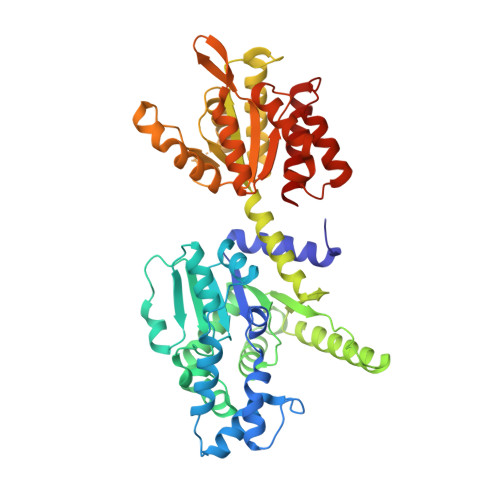
Activation of Thoeris antiviral system via SIR2 effector filament assembly.

Tamulaitiene, G.Sabonis, D.Sasnauskas, G.Ruksenaite, A.Silanskas, A.Avraham, C.Ofir, G.Sorek, R.Zaremba, M.Siksnys, V.

(2024) Nature 627: 431-436

  • DOI: https://doi.org/10.1038/s41586-024-07092-x
  • Primary Citation Related Structures: 
    8BTN, 8BTO, 8BTP

  • PubMed Abstract: 

    To survive bacteriophage (phage) infections, bacteria developed numerous anti-phage defence systems 1-7 . Some of them (for example, type III CRISPR-Cas, CBASS, Pycsar and Thoeris) consist of two modules: a sensor responsible for infection recognition and an effector that stops viral replication by destroying key cellular components 8-12 . In the Thoeris system, a Toll/interleukin-1 receptor (TIR)-domain protein, ThsB, acts as a sensor that synthesizes an isomer of cyclic ADP ribose, 1''-3' glycocyclic ADP ribose (gcADPR), which is bound in the Smf/DprA-LOG (SLOG) domain of the ThsA effector and activates the silent information regulator 2 (SIR2)-domain-mediated hydrolysis of a key cell metabolite, NAD + (refs.  12-14 ). Although the structure of ThsA has been solved 15 , the ThsA activation mechanism remained incompletely understood. Here we show that 1''-3' gcADPR, synthesized in vitro by the dimeric ThsB' protein, binds to the ThsA SLOG domain, thereby activating ThsA by triggering helical filament assembly of ThsA tetramers. The cryogenic electron microscopy (cryo-EM) structure of activated ThsA revealed that filament assembly stabilizes the active conformation of the ThsA SIR2 domain, enabling rapid NAD + depletion. Furthermore, we demonstrate that filament formation enables a switch-like response of ThsA to the 1''-3' gcADPR signal.


  • Organizational Affiliation
    • Institute of Biotechnology, Life Sciences Center, Vilnius University, Vilnius, Lithuania. giedre.tamulaitiene@bti.vu.lt.

Macromolecule Content 

  • Total Structure Weight: 679.47 kDa 
  • Atom Count: 46,128 
  • Modeled Residue Count: 5,676 
  • Deposited Residue Count: 5,748 
  • Unique protein chains: 1

Macromolecules

Find similar proteins by:|  3D Structure
Entity ID: 1
MoleculeChains  Sequence LengthOrganismDetailsImage
NAD(+) hydrolase ThsA
A, B, C, D, E
A, B, C, D, E, F, G, H, I, J, K, L
479Bacillus cereus MSX-D12Mutation(s): 1 
Gene Names: thsAII9_05448
EC: 3.2.2.5
UniProt
Find proteins for J8G6Z1 (Bacillus cereus (strain MSX-D12))
Explore J8G6Z1 
Go to UniProtKB:  J8G6Z1
Entity Groups
Sequence Clusters30% Identity50% Identity70% Identity90% Identity95% Identity100% Identity
UniProt GroupJ8G6Z1
Sequence Annotations
Expand
Reference Sequence

Small Molecules

Ligands 2 Unique
IDChains Name / Formula / InChI Key2D Diagram3D Interactions
NAD
(Subject of Investigation/LOI)

Query on NAD


Download:Ideal Coordinates CCD File
BA [auth H]
DA [auth I]
FA [auth J]
HA [auth K]
JA [auth L]
BA [auth H],
DA [auth I],
FA [auth J],
HA [auth K],
JA [auth L],
N [auth A],
P [auth B],
R [auth C],
T [auth D],
V [auth E],
X [auth F],
Z [auth G]
NICOTINAMIDE-ADENINE-DINUCLEOTIDE
C21 H27 N7 O14 P2
BAWFJGJZGIEFAR-NNYOXOHSSA-N
OJC
(Subject of Investigation/LOI)

Query on OJC


Download:Ideal Coordinates CCD File
AA [auth H]
CA [auth I]
EA [auth J]
GA [auth K]
IA [auth L]
AA [auth H],
CA [auth I],
EA [auth J],
GA [auth K],
IA [auth L],
M [auth A],
O [auth B],
Q [auth C],
S [auth D],
U [auth E],
W [auth F],
Y [auth G]
(2R,3R,3aS,5S,6R,7S,8R,11R,13S,15aR)-2-(6-amino-9H-purin-9-yl)-3,6,7,11,13-pentahydroxyoctahydro-2H,5H,11H,13H-5,8-epoxy-11lambda~5~,13lambda~5~-furo[2,3-g][1,3,5,9,2,4]tetraoxadiphosphacyclotetradecine-11,13-dione
C15 H21 N5 O13 P2
NDSLKXAJVDJCEG-ZQSHOCFMSA-N

Experimental Data & Validation

Experimental Data

  • Method: ELECTRON MICROSCOPY
  • Resolution: 2.96 Å
  • Aggregation State: HELICAL ARRAY 
  • Reconstruction Method: HELICAL 
EM Software:
TaskSoftware PackageVersion
MODEL REFINEMENTPHENIX
RECONSTRUCTIONcryoSPARC

Structure Validation

View Full Validation Report


Entry History 

& Funding Information

Deposition Data


Funding OrganizationLocationGrant Number
Research Council of LithuaniaLithuaniaS-MIP-21-6

Revision History  (Full details and data files)

  • Version 1.0: 2024-02-21
    Type: Initial release
  • Version 1.1: 2024-03-06
    Changes: Database references
  • Version 1.2: 2024-03-27
    Changes: Database references
  • Version 1.3: 2024-04-03
    Changes: Database references