8BSE | pdb_00008bse

CRYSTAL STRUCTURE OF SARS-COV-2 RECEPTOR BINDING DOMAIN (RBD) in complex with 1D1 Fab


Experimental Data Snapshot

  • Method: X-RAY DIFFRACTION
  • Resolution: 1.90 Å
  • R-Value Free: 
    0.216 (Depositor), 0.225 (DCC) 
  • R-Value Work: 
    0.177 (Depositor), 0.189 (DCC) 
  • R-Value Observed: 
    0.179 (Depositor) 

Starting Models: experimental
View more details

wwPDB Validation 3D Report Full Report

Validation slider image for 8BSE

Ligand Structure Quality Assessment 


This is version 1.2 of the entry. See complete history

Literature

Efficacy of the combination of monoclonal antibodies against the SARS-CoV-2 Beta and Delta variants.

Boonkrai, C.Cotrone, T.S.Chaisuriyong, W.Tantawichien, T.Thisyakorn, U.Fernandez, S.Hunsawong, T.Reed, M.Wongtangprasert, T.Audomsun, T.Phakham, T.Attakitbancha, C.Saelao, P.Focht, D.Kimbung, R.Welin, M.Malik, A.A.Pisitkun, T.Srisawat, N.

(2023) PLoS One 18: e0284173-e0284173

  • DOI: https://doi.org/10.1371/journal.pone.0284173
  • Primary Citation Related Structures: 
    8BSE, 8BSF

  • PubMed Abstract: 

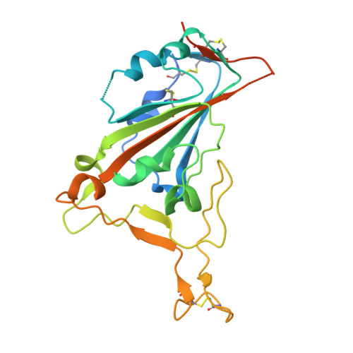
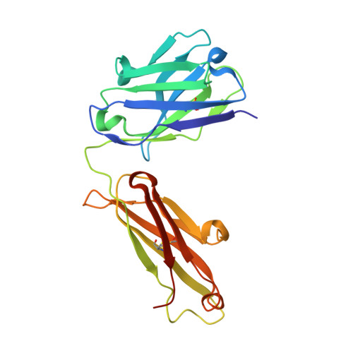
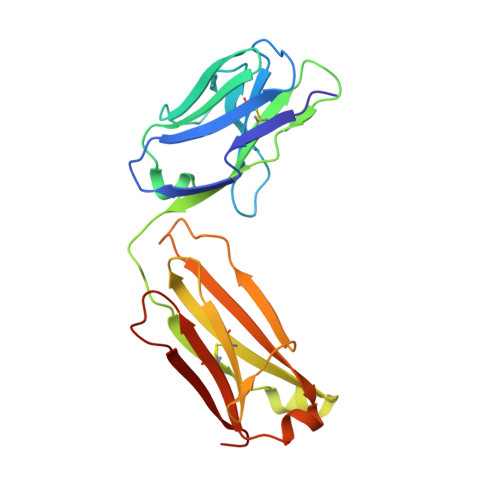
    The pandemic of severe acute respiratory syndrome coronavirus 2 (SARS-CoV-2) is currently the biggest healthcare issue worldwide. This study aimed to develop a monoclonal antibody against SARS-CoV-2 from B cells of recovered COVID-19 patients, which might have beneficial therapeutic purposes for COVID-19 patients. We successfully generated human monoclonal antibodies (hmAbs) against the receptor binding domain (RBD) protein of SARS-CoV-2 using developed hybridoma technology. The isolated hmAbs against the RBD protein (wild-type) showed high binding activity and neutralized the interaction between the RBD and the cellular receptor angiotensin-converting enzyme 2 (ACE2) protein. Epitope binning and crystallography results displayed target epitopes of these antibodies in distinct regions beneficial in the mix as a cocktail. The 3D2 binds to conserved epitopes among multi-variants. Pseudovirion-based neutralization results revealed that the antibody cocktail, 1D1 and 3D2, showed high potency in multiple variants of SARS-CoV-2 infection. In vivo studies showed the ability of the antibody cocktail treatment (intraperitoneal (i.p.) administration) to reduce viral load (Beta variant) in blood and various tissues. While the antibody cocktail treatment (intranasal (i.n.) administration) could not significantly reduce the viral load in nasal turbinate and lung tissue, it could reduce the viral load in blood, kidney, and brain tissue. These findings revealed that the efficacy of the antibody cocktail, 1D1 and 3D2, should be further studied in animal models in terms of timing of administration, optimal dose, and efficacy to mitigate inflammation in targeted tissue such as nasal turbinate and lung.


  • Organizational Affiliation
    • Interdisciplinary Program of Biomedical Sciences, Graduate School, Chulalongkorn University, Bangkok, Thailand.

Macromolecule Content 

  • Total Structure Weight: 73.67 kDa 
  • Atom Count: 5,349 
  • Modeled Residue Count: 619 
  • Deposited Residue Count: 666 
  • Unique protein chains: 3

Macromolecules

Find similar proteins by:|  3D Structure
Entity ID: 1
MoleculeChains  Sequence LengthOrganismDetailsImage
Spike glycoprotein231Severe acute respiratory syndrome coronavirus 2Mutation(s): 0 
Gene Names: S2
UniProt
Find proteins for P0DTC2 (Severe acute respiratory syndrome coronavirus 2)
Explore P0DTC2 
Go to UniProtKB:  P0DTC2
Entity Groups
Sequence Clusters30% Identity50% Identity70% Identity90% Identity95% Identity100% Identity
UniProt GroupP0DTC2
Glycosylation
Glycosylation Sites: 1Go to GlyGen: P0DTC2-1
Sequence Annotations
Expand
Reference Sequence
Find similar proteins by:|  3D Structure
Entity ID: 2
MoleculeChains  Sequence LengthOrganismDetailsImage
1D1 FAB HEAVY CHAINB [auth H]221Homo sapiensMutation(s): 0 
Find similar proteins by:|  3D Structure
Entity ID: 3
MoleculeChains  Sequence LengthOrganismDetailsImage
1D1 FAB LIGHT CHAINC [auth L]214Homo sapiensMutation(s): 0 

Small Molecules

Ligands 3 Unique
IDChains Name / Formula / InChI Key2D Diagram3D Interactions
NAG

Query on NAG


Download:Ideal Coordinates CCD File
D [auth A]2-acetamido-2-deoxy-beta-D-glucopyranose
C8 H15 N O6
OVRNDRQMDRJTHS-FMDGEEDCSA-N
TRS

Query on TRS


Download:Ideal Coordinates CCD File
I [auth H]2-AMINO-2-HYDROXYMETHYL-PROPANE-1,3-DIOL
C4 H12 N O3
LENZDBCJOHFCAS-UHFFFAOYSA-O
GOL

Query on GOL


Download:Ideal Coordinates CCD File
E [auth A],
F [auth H],
G [auth H],
H,
J [auth L]
GLYCEROL
C3 H8 O3
PEDCQBHIVMGVHV-UHFFFAOYSA-N

Experimental Data & Validation

Experimental Data

  • Method: X-RAY DIFFRACTION
  • Resolution: 1.90 Å
  • R-Value Free:  0.216 (Depositor), 0.225 (DCC) 
  • R-Value Work:  0.177 (Depositor), 0.189 (DCC) 
  • R-Value Observed: 0.179 (Depositor) 
Space Group: P 21 21 21
Unit Cell:
Length ( Å )Angle ( ˚ )
a = 56.177α = 90
b = 110.018β = 90
c = 149.392γ = 90
Software Package:
Software NamePurpose
REFMACrefinement
Aimlessdata scaling
PHASERphasing
XDSdata reduction

Structure Validation

View Full Validation Report


Ligand Structure Quality Assessment 


Entry History 

& Funding Information

Deposition Data


Funding OrganizationLocationGrant Number
Other governmentThailand--

Revision History  (Full details and data files)

  • Version 1.0: 2023-05-17
    Type: Initial release
  • Version 1.1: 2023-10-18
    Changes: Data collection, Database references, Source and taxonomy
  • Version 1.2: 2024-10-09
    Changes: Structure summary