8BS9 | pdb_00008bs9

Structure of USP36 in complex with Ubiquitin-PA


Experimental Data Snapshot

  • Method: X-RAY DIFFRACTION
  • Resolution: 1.90 Å
  • R-Value Free: 
    0.209 (Depositor), 0.205 (DCC) 
  • R-Value Work: 
    0.171 (Depositor), 0.167 (DCC) 
  • R-Value Observed: 
    0.173 (Depositor) 

Starting Models: in silico, experimental
View more details

wwPDB Validation 3D Report Full Report

Validation slider image for 8BS9

Ligand Structure Quality Assessment 


This is version 1.3 of the entry. See complete history

Literature

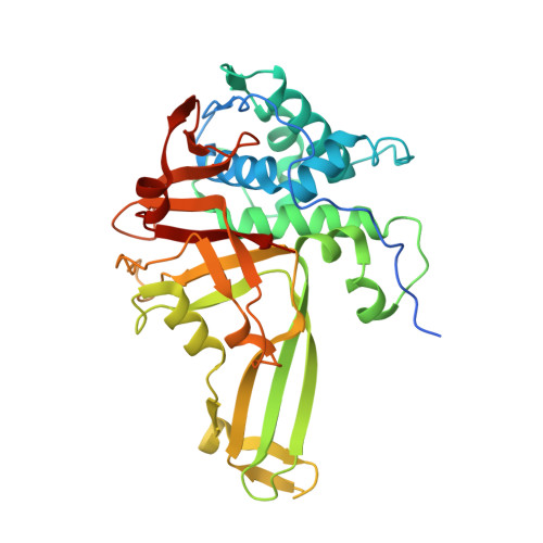
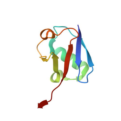
Molecular basis for ubiquitin/Fubi cross-reactivity in USP16 and USP36.

O'Dea, R.Kazi, N.Hoffmann-Benito, A.Zhao, Z.Recknagel, S.Wendrich, K.Janning, P.Gersch, M.

(2023) Nat Chem Biol 19: 1394-1405

  • DOI: https://doi.org/10.1038/s41589-023-01388-1
  • Primary Citation Related Structures: 
    8BS3, 8BS9

  • PubMed Abstract: 

    Ubiquitin and ubiquitin-like proteins typically use distinct machineries to facilitate diverse functions. The immunosuppressive ubiquitin-like protein Fubi is synthesized as an N-terminal fusion to a ribosomal protein (Fubi-S30). Its proteolytic maturation by the nucleolar deubiquitinase USP36 is strictly required for translationally competent ribosomes. What endows USP36 with this activity, how Fubi is recognized and whether other Fubi proteases exist are unclear. Here, we report a chemical tool kit that facilitated the discovery of dual ubiquitin/Fubi cleavage activity in USP16 in addition to USP36 by chemoproteomics. Crystal structures of USP36 complexed with Fubi and ubiquitin uncover its substrate recognition mechanism and explain how other deubiquitinases are restricted from Fubi. Furthermore, we introduce Fubi C-terminal hydrolase measurements and reveal a synergistic role of USP16 in Fubi-S30 maturation. Our data highlight how ubiquitin/Fubi specificity is achieved in a subset of human deubiquitinases and open the door to a systematic investigation of the Fubi system.


  • Organizational Affiliation
    • Chemical Genomics Centre, Max Planck Institute of Molecular Physiology, Dortmund, Germany.

Macromolecule Content 

  • Total Structure Weight: 95.53 kDa 
  • Atom Count: 6,874 
  • Modeled Residue Count: 798 
  • Deposited Residue Count: 842 
  • Unique protein chains: 2

Macromolecules

Find similar proteins by:|  3D Structure
Entity ID: 1
MoleculeChains  Sequence LengthOrganismDetailsImage
Ubiquitin carboxyl-terminal hydrolase 36
A, C
346Homo sapiensMutation(s): 0 
Gene Names: USP36KIAA1453
EC: 3.4.19.12 (PDB Primary Data), 2.3.2 (UniProt)
UniProt & NIH Common Fund Data Resources
Find proteins for Q9P275 (Homo sapiens)
Explore Q9P275 
Go to UniProtKB:  Q9P275
PHAROS:  Q9P275
GTEx:  ENSG00000055483 
Entity Groups
Sequence Clusters30% Identity50% Identity70% Identity90% Identity95% Identity100% Identity
UniProt GroupQ9P275
Sequence Annotations
Expand
Reference Sequence
Find similar proteins by:|  3D Structure
Entity ID: 2
MoleculeChains  Sequence LengthOrganismDetailsImage
Polyubiquitin-B
B, D
75Homo sapiensMutation(s): 0 
Gene Names: UBB
UniProt & NIH Common Fund Data Resources
Find proteins for P0CG47 (Homo sapiens)
Explore P0CG47 
Go to UniProtKB:  P0CG47
PHAROS:  P0CG47
GTEx:  ENSG00000170315 
Entity Groups
Sequence Clusters30% Identity50% Identity70% Identity90% Identity95% Identity100% Identity
UniProt GroupP0CG47
Sequence Annotations
Expand
Reference Sequence

Small Molecules

Ligands 3 Unique
IDChains Name / Formula / InChI Key2D Diagram3D Interactions
ZN

Query on ZN


Download:Ideal Coordinates CCD File
E [auth A],
F [auth A],
I [auth C],
J [auth C]
ZINC ION
Zn
PTFCDOFLOPIGGS-UHFFFAOYSA-N
AYE
(Subject of Investigation/LOI)

Query on AYE


Download:Ideal Coordinates CCD File
G [auth A],
K [auth C]
prop-2-en-1-amine
C3 H7 N
VVJKKWFAADXIJK-UHFFFAOYSA-N
NA

Query on NA


Download:Ideal Coordinates CCD File
H [auth A],
L [auth C]
SODIUM ION
Na
FKNQFGJONOIPTF-UHFFFAOYSA-N

Experimental Data & Validation

Experimental Data

  • Method: X-RAY DIFFRACTION
  • Resolution: 1.90 Å
  • R-Value Free:  0.209 (Depositor), 0.205 (DCC) 
  • R-Value Work:  0.171 (Depositor), 0.167 (DCC) 
  • R-Value Observed: 0.173 (Depositor) 
Space Group: P 1 21 1
Unit Cell:
Length ( Å )Angle ( ˚ )
a = 75.743α = 90
b = 65.307β = 93.265
c = 103.411γ = 90
Software Package:
Software NamePurpose
PHENIXrefinement
XDSdata reduction
Aimlessdata scaling
PHASERphasing
Cootmodel building

Structure Validation

View Full Validation Report


Ligand Structure Quality Assessment 


Entry History 

& Funding Information

Deposition Data


Funding OrganizationLocationGrant Number
Max Planck SocietyGermanyCGC-III-352S

Revision History  (Full details and data files)

  • Version 1.0: 2023-07-12
    Type: Initial release
  • Version 1.1: 2023-07-26
    Changes: Database references
  • Version 1.2: 2023-11-08
    Changes: Data collection, Database references
  • Version 1.3: 2023-11-15
    Changes: Database references