8BRH | pdb_00008brh

Co-crystal structure of She4 with Myo4 peptide


Experimental Data Snapshot

  • Method: X-RAY DIFFRACTION
  • Resolution: 2.40 Å
  • R-Value Free: 
    0.250 (Depositor), 0.250 (DCC) 
  • R-Value Work: 
    0.212 (Depositor), 0.212 (DCC) 
  • R-Value Observed: 
    0.214 (Depositor) 

Starting Model: experimental
View more details

wwPDB Validation 3D Report Full Report

Validation slider image for 8BRH

This is version 1.1 of the entry. See complete history

Literature

UNC-45 assisted myosin folding depends on a conserved FX 3 HY motif implicated in Freeman Sheldon Syndrome.

Vogel, A.Arnese, R.Gudino Carrillo, R.M.Sehr, D.Deszcz, L.Bylicki, A.Meinhart, A.Clausen, T.

(2024) Nat Commun 15: 6272-6272

  • DOI: https://doi.org/10.1038/s41467-024-50442-6
  • Primary Citation Related Structures: 
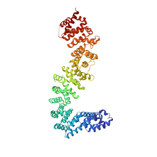
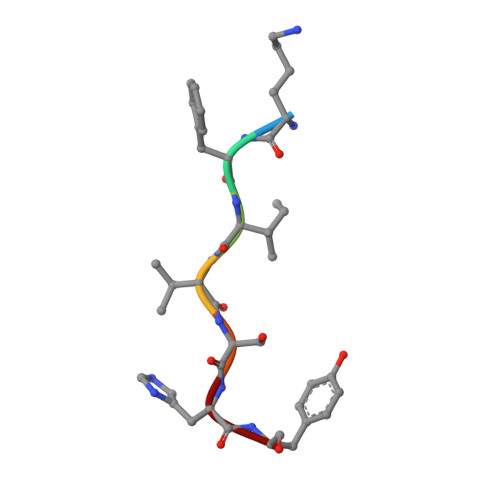
    8BRG, 8BRH

  • PubMed Abstract: 

    Myosin motors are critical for diverse motility functions, ranging from cytokinesis and endocytosis to muscle contraction. The UNC-45 chaperone controls myosin function mediating the folding, assembly, and degradation of the muscle protein. Here, we analyze the molecular mechanism of UNC-45 as a hub in myosin quality control. We show that UNC-45 forms discrete complexes with folded and unfolded myosin, forwarding them to downstream chaperones and E3 ligases. Structural analysis of a minimal chaperone:substrate complex reveals that UNC-45 binds to a conserved FX 3 HY motif in the myosin motor domain. Disrupting the observed interface by mutagenesis prevents myosin maturation leading to protein aggregation in vivo. We also show that a mutation in the FX 3 HY motif linked to the Freeman Sheldon Syndrome impairs UNC-45 assisted folding, reducing the level of functional myosin. These findings demonstrate that a faulty myosin quality control is a critical yet unexplored cause of human myopathies.


  • Organizational Affiliation
    • Research Institute of Molecular Pathology, Vienna BioCenter, Vienna, Austria.

Macromolecule Content 

  • Total Structure Weight: 76.75 kDa 
  • Atom Count: 5,215 
  • Modeled Residue Count: 639 
  • Deposited Residue Count: 676 
  • Unique protein chains: 2

Macromolecules

Find similar proteins by:|  3D Structure
Entity ID: 1
MoleculeChains  Sequence LengthOrganismDetailsImage
KLLA0E16699p669Kluyveromyces lactisMutation(s): 0 
Gene Names: KLLA0_E16699g
UniProt
Find proteins for Q6CMY7 (Kluyveromyces lactis (strain ATCC 8585 / CBS 2359 / DSM 70799 / NBRC 1267 / NRRL Y-1140 / WM37))
Explore Q6CMY7 
Go to UniProtKB:  Q6CMY7
Entity Groups
Sequence Clusters30% Identity50% Identity70% Identity90% Identity95% Identity100% Identity
UniProt GroupQ6CMY7
Sequence Annotations
Expand
Reference Sequence
Find similar proteins by:  Sequence   |   3D Structure  
Entity ID: 2
MoleculeChains  Sequence LengthOrganismDetailsImage
Myo4 peptide (LYS-PHE-ILE-VAL-SER-HIS-TYR)7Saccharomyces cerevisiaeMutation(s): 0 
UniProt
Find proteins for P19524 (Saccharomyces cerevisiae (strain ATCC 204508 / S288c))
Explore P19524 
Go to UniProtKB:  P19524
Entity Groups
UniProt GroupP19524
Sequence Annotations
Expand
Reference Sequence

Experimental Data & Validation

Experimental Data

  • Method: X-RAY DIFFRACTION
  • Resolution: 2.40 Å
  • R-Value Free:  0.250 (Depositor), 0.250 (DCC) 
  • R-Value Work:  0.212 (Depositor), 0.212 (DCC) 
  • R-Value Observed: 0.214 (Depositor) 
Space Group: P 31 2 1
Unit Cell:
Length ( Å )Angle ( ˚ )
a = 128.48α = 90
b = 128.48β = 90
c = 77.54γ = 120
Software Package:
Software NamePurpose
PHENIXrefinement
XDSdata reduction
XSCALEdata scaling
PHASERphasing

Structure Validation

View Full Validation Report


Entry History 

& Funding Information

Deposition Data


Funding OrganizationLocationGrant Number
Austrian Science FundAustria--

Revision History  (Full details and data files)

  • Version 1.0: 2024-06-05
    Type: Initial release
  • Version 1.1: 2024-08-14
    Changes: Database references