


Crystal structure of Trichoplax Dlg PDZ3 domain
Maddumage, J.C., Kvansakul, M., Humbert, P.O.To be published.
Experimental Data Snapshot
Starting Model: experimental
View more details
wwPDB Validation 3D Report Full Report

Entity ID: 1 | |||||
|---|---|---|---|---|---|
| Molecule | Chains | Sequence Length | Organism | Details | Image |
| Disks large-like protein 1 | 93 | Trichoplax sp. H2 | Mutation(s): 0 Gene Names: TrispH2_000924 | 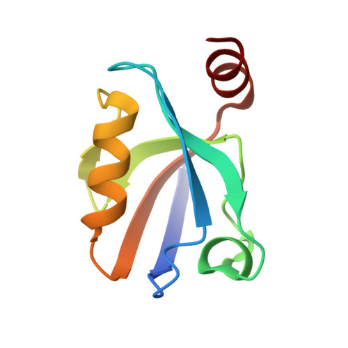 | |
UniProt | |||||
Entity Groups | |||||
| Sequence Clusters | 30% Identity50% Identity70% Identity90% Identity95% Identity100% Identity | ||||
| UniProt Group | A0ACD6BAH5 | ||||
Sequence AnnotationsExpand | |||||
Reference Sequence | |||||
| Length ( Å ) | Angle ( ˚ ) |
|---|---|
| a = 32.375 | α = 90 |
| b = 35.434 | β = 90 |
| c = 72.99 | γ = 90 |
| Software Name | Purpose |
|---|---|
| PHENIX | refinement |
| DIALS | data reduction |
| Aimless | data scaling |
| PHASER | phasing |
| Funding Organization | Location | Grant Number |
|---|---|---|
| National Health and Medical Research Council (NHMRC, Australia) | Australia | APP1103871 |
| Australian Research Council (ARC) | Australia | FT130101349 |


