8BPG

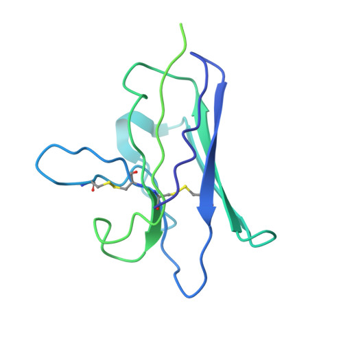
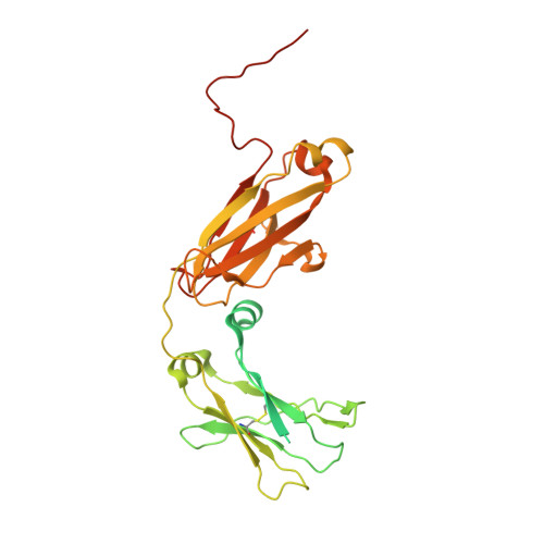
FcMR binding at subunit Fcu3 of IgM pentamer


Experimental Data Snapshot

  • Method: ELECTRON MICROSCOPY
  • Resolution: 3.10 Å
  • Aggregation State: PARTICLE 
  • Reconstruction Method: SINGLE PARTICLE 

wwPDB Validation   3D Report Full Report


This is version 1.4 of the entry. See complete history


Literature

Structural basis for Fc receptor recognition of immunoglobulin M.

Chen, Q.Menon, R.P.Masino, L.Tolar, P.Rosenthal, P.B.

(2023) Nat Struct Mol Biol 30: 1033-1039

  • DOI: https://doi.org/10.1038/s41594-023-00985-x
  • Primary Citation of Related Structures:  
    8BPE, 8BPF, 8BPG

  • PubMed Abstract: 

    Immunoglobulin Fc receptors are cell surface transmembrane proteins that bind to the Fc constant region of antibodies and play critical roles in regulating immune responses by activation of immune cells, clearance of immune complexes and regulation of antibody production. FcμR is the immunoglobulin M (IgM) antibody isotype-specific Fc receptor involved in the survival and activation of B cells. Here we reveal eight binding sites for the human FcμR immunoglobulin domain on the IgM pentamer by cryogenic electron microscopy. One of the sites overlaps with the binding site for the polymeric immunoglobulin receptor (pIgR), but a different mode of FcμR binding explains its antibody isotype specificity. Variation in FcμR binding sites and their occupancy reflects the asymmetry of the IgM pentameric core and the versatility of FcμR binding. The complex explains engagement with polymeric serum IgM and the monomeric IgM B-cell receptor (BCR).


  • Organizational Affiliation

    Structural Biology Science Technology Platform, The Francis Crick Institute, London, UK.


Macromolecules
Find similar proteins by:  (by identity cutoff)  |  3D Structure
Entity ID: 1
MoleculeChains Sequence LengthOrganismDetailsImage
Fas apoptotic inhibitory molecule 3A [auth B],
D [auth A]
234Homo sapiensMutation(s): 0 
Gene Names: FCMRFAIM3TOSO
UniProt & NIH Common Fund Data Resources
Find proteins for O60667 (Homo sapiens)
Go to UniProtKB:  O60667
PHAROS:  O60667
GTEx:  ENSG00000162894 
Sequence Annotations
Expand
  • Reference Sequence
Find similar proteins by:  (by identity cutoff)  |  3D Structure
Entity ID: 2
MoleculeChains Sequence LengthOrganismDetailsImage
Immunoglobulin heavy constant muB [auth D],
C,
E,
F
348Homo sapiensMutation(s): 0 
UniProt & NIH Common Fund Data Resources
Find proteins for P01871 (Homo sapiens)
Explore P01871 
Go to UniProtKB:  P01871
PHAROS:  P01871
GTEx:  ENSG00000211899 
Entity Groups  
Sequence Clusters30% Identity50% Identity70% Identity90% Identity95% Identity100% Identity
UniProt GroupP01871
Glycosylation
Glycosylation Sites: 1Go to GlyGen: P01871-2
Sequence Annotations
Expand
  • Reference Sequence
Oligosaccharides

Help

Entity ID: 3
MoleculeChains Length2D Diagram Glycosylation3D Interactions
2-acetamido-2-deoxy-beta-D-glucopyranose-(1-4)-2-acetamido-2-deoxy-beta-D-glucopyranose
G, H
2N-Glycosylation
Glycosylation Resources
GlyTouCan:  G42666HT
GlyCosmos:  G42666HT
GlyGen:  G42666HT
Experimental Data & Validation

Experimental Data

  • Method: ELECTRON MICROSCOPY
  • Resolution: 3.10 Å
  • Aggregation State: PARTICLE 
  • Reconstruction Method: SINGLE PARTICLE 

Structure Validation

View Full Validation Report


Entry History & Funding Information

Deposition Data


Funding OrganizationLocationGrant Number
Cancer Research UKUnited KingdomFC001143
Medical Research Council (MRC, United Kingdom)United KingdomFC001143
Wellcome TrustUnited KingdomFC001143
Cancer Research UKUnited KingdomFC001185
Medical Research Council (MRC, United Kingdom)United KingdomFC001185
Wellcome TrustUnited KingdomFC001185

Revision History  (Full details and data files)

  • Version 1.0: 2023-04-12
    Type: Initial release
  • Version 1.1: 2023-04-26
    Changes: Database references
  • Version 1.2: 2023-05-03
    Changes: Database references
  • Version 1.3: 2023-07-26
    Changes: Database references
  • Version 1.4: 2024-10-16
    Changes: Data collection, Structure summary