8BP6 | pdb_00008bp6

Structure of MHC-class I related molecule MR1 with bound M3Ade.


Experimental Data Snapshot

  • Method: X-RAY DIFFRACTION
  • Resolution: 2.80 Å
  • R-Value Free: 
    0.247 (Depositor), 0.240 (DCC) 
  • R-Value Work: 
    0.237 (Depositor), 0.235 (DCC) 
  • R-Value Observed: 
    0.236 (Depositor) 

Starting Model: experimental
View more details

wwPDB Validation 3D Report Full Report

Validation slider image for 8BP6

Ligand Structure Quality Assessment 


This is version 1.4 of the entry. See complete history

Literature

The carbonyl nucleobase adduct M 3 Ade is a potent antigen for adaptive polyclonal MR1-restricted T cells.

Chancellor, A.Constantin, D.Berloffa, G.Yang, Q.Nosi, V.Loureiro, J.P.Colombo, R.Jakob, R.P.Joss, D.Pfeffer, M.De Simone, G.Morabito, A.Schaefer, V.Vacchini, A.Brunelli, L.Montagna, D.Heim, M.Zippelius, A.Davoli, E.Haussinger, D.Maier, T.Mori, L.De Libero, G.

(2024) Immunity 

  • DOI: https://doi.org/10.1016/j.immuni.2024.11.019
  • Primary Citation Related Structures: 
    8BP6

  • PubMed Abstract: 

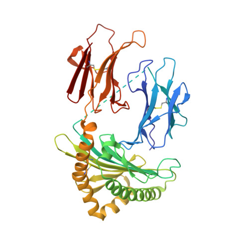
    The major histocompatibility complex (MHC) class I-related molecule MHC-class-I-related protein 1 (MR1) presents metabolites to distinct MR1-restricted T cell subsets, including mucosal-associated invariant T (MAIT) and MR1T cells. However, self-reactive MR1T cells and the nature of recognized antigens remain underexplored. Here, we report a cell endogenous carbonyl adduct of adenine (8-(9H-purin-6-yl)-2-oxa-8-azabicyclo[3.3.1]nona-3,6-diene-4,6-dicarbaldehyde [M 3 Ade]) sequestered in the A' pocket of MR1. M 3 Ade induced in vitro MR1-mediated stimulation of MR1T cell clones that bound MR1-M 3 Ade tetramers. MR1-M 3 Ade tetramers identified heterogeneous MR1-reactive T cells ex vivo in healthy donors, individuals with acute myeloid leukemia, and tumor-infiltrating lymphocytes from non-small cell lung adenocarcinoma and hepatocarcinoma. These cells displayed phenotypic, transcriptional, and functional diversity at distinct differentiation stages, indicating their adaptive nature. They were also polyclonal, with some preferential T cell receptor (TCRαβ) pair usage. Thus, M 3 Ade is an MR1-presented self-metabolite that enables stimulation and tracking of human-MR1T cells from blood and tissue, aiding our understanding of their roles in health and disease.


  • Organizational Affiliation
    • Experimental Immunology, Department of Biomedicine, University Hospital Basel, University of Basel, 4031 Basel, Switzerland. Electronic address: andrew.chancellor@unibas.ch.

Macromolecule Content 

  • Total Structure Weight: 89.2 kDa 
  • Atom Count: 6,156 
  • Modeled Residue Count: 737 
  • Deposited Residue Count: 768 
  • Unique protein chains: 1

Macromolecules

Find similar proteins by:|  3D Structure
Entity ID: 1
MoleculeChains  Sequence LengthOrganismDetailsImage
Beta-2-microglobulin,Major histocompatibility complex class I-related gene protein
A, B
384Homo sapiensMutation(s): 0 
Gene Names: B2MMR1
UniProt & NIH Common Fund Data Resources
Find proteins for P61769 (Homo sapiens)
Explore P61769 
Go to UniProtKB:  P61769
PHAROS:  P61769
GTEx:  ENSG00000166710 
Find proteins for Q95460 (Homo sapiens)
Explore Q95460 
Go to UniProtKB:  Q95460
PHAROS:  Q95460
GTEx:  ENSG00000153029 
Entity Groups
Sequence Clusters30% Identity50% Identity70% Identity90% Identity95% Identity100% Identity
UniProt GroupsQ95460P61769
Sequence Annotations
Expand
Reference Sequence

Small Molecules

Ligands 1 Unique
IDChains Name / Formula / InChI Key2D Diagram3D Interactions
R8F
(Subject of Investigation/LOI)

Query on R8F


Download:Ideal Coordinates CCD File
C [auth A],
D [auth B]
(1R,5S)-8-(9H-purin-6-yl)-2-oxa-8-azabicyclo[3.3.1]nona-3,6-diene-4,6-dicarbaldehyde
C14 H11 N5 O3
JDSDLPGCRCFXJZ-WDEREUQCSA-N

Experimental Data & Validation

Experimental Data

  • Method: X-RAY DIFFRACTION
  • Resolution: 2.80 Å
  • R-Value Free:  0.247 (Depositor), 0.240 (DCC) 
  • R-Value Work:  0.237 (Depositor), 0.235 (DCC) 
  • R-Value Observed: 0.236 (Depositor) 
Space Group: P 1 21 1
Unit Cell:
Length ( Å )Angle ( ˚ )
a = 50.352α = 90
b = 143.534β = 101.82
c = 77.029γ = 90
Software Package:
Software NamePurpose
PHENIXrefinement
XDSdata reduction
Aimlessdata scaling
PHASERphasing

Structure Validation

View Full Validation Report


Ligand Structure Quality Assessment 


Entry History 

& Funding Information

Deposition Data


Funding OrganizationLocationGrant Number
Swiss National Science FoundationSwitzerland--

Revision History  (Full details and data files)

  • Version 1.0: 2023-11-29
    Type: Initial release
  • Version 1.1: 2024-10-23
    Changes: Structure summary
  • Version 1.2: 2025-01-08
    Changes: Database references
  • Version 1.3: 2025-02-05
    Changes: Database references, Structure summary
  • Version 1.4: 2026-03-04
    Changes: Refinement description