A Complementarity-Based Approach to De Novo Binder Design.
Maksymenko, K., Hatskovska, V., Coles, M., Aghaallaei, N., Pashkovskaia, N., Borbaran-Bravo, N., Pilz, M., Bucher, P., Volz, M., Pereira, J., Hartmann, M.D., Tabatabai, G., Feucht, J., Liebau, S., Muller, P., Lupas, A.N., Skokowa, J., ElGamacy, M.(2025) Adv Sci (Weinh) 12: e02015-e02015
- PubMed: 40686280 Search on PubMedSearch on PubMed Central
- DOI: https://doi.org/10.1002/advs.202502015
- Primary Citation Related Structures:
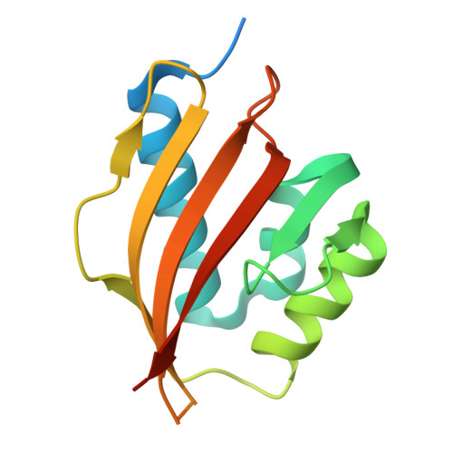
8BL5, 8BL9 - PubMed Abstract:
De novo design of binders capable of targeting arbitrarily selected epitopes remains a substantial challenge. Here, a generalizable computational strategy is presented to design site-specific protein binders, obviating steps of extensive empirical optimization or in vitro screening. The dock-and-design pipeline retrieves complementary scaffolds from a protein structure database to a given query epitope, where the scaffold is mutated to carve a binding site de novo. The docking step utilizes a novel fingerprint that greatly simplifies and accelerates the surface complementarity evaluation. As proof-of-concept, we designed protein binders to target three distinct epitopes on two different oncogenic targets; vascular endothelial growth factor (VEGF) and interleukin-7 receptor-α (IL-7Rα). Experimental characterization of only 24 candidates identified nanomolar binders against each of the target epitopes, where the binders belonged to five different folds. Several designs were active in vitro. Moreover, anti-VEGF designs showed tumor-inhibiting activity in vivo, highlighting their therapeutic potential.
- Department of Protein Evolution, Max Planck Institute for Biology, 72076, Tübingen, Germany.
Organizational Affiliation: