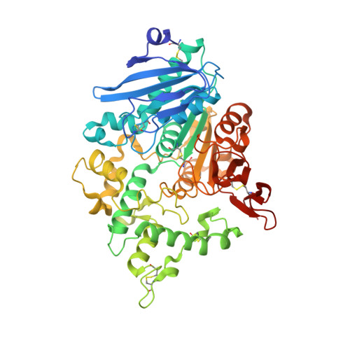
8BHH

The crystal structure of a feruloyl esterase C from Fusarium oxysporum in complex with p-coumaric acid


Experimental Data Snapshot

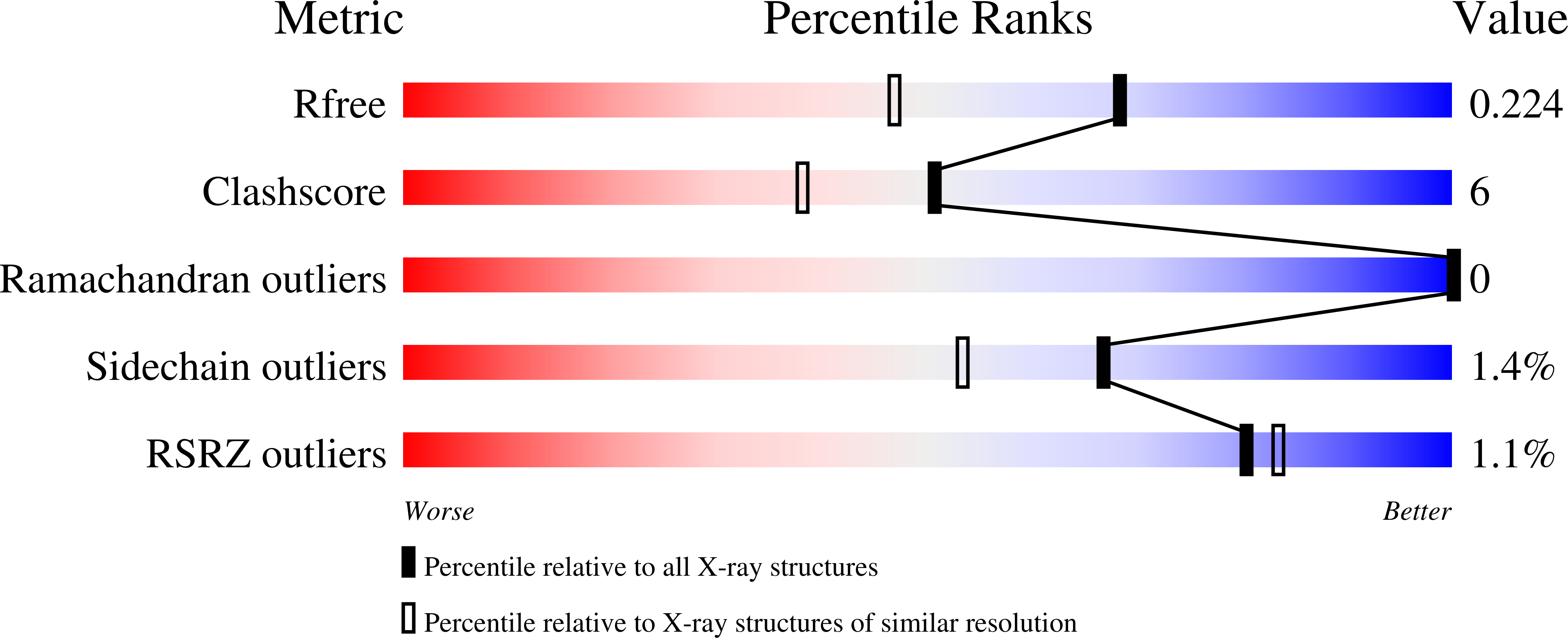
  • Method: X-RAY DIFFRACTION
  • Resolution: 1.69 Å
  • R-Value Free: 0.217 
  • R-Value Work: 0.171 
  • R-Value Observed: 0.173 

Starting Model: experimental
View more details

wwPDB Validation   3D Report Full Report


Ligand Structure Quality Assessment 


This is version 1.2 of the entry. See complete history


Literature

Crystal structure of the Fusarium oxysporum tannase-like feruloyl esterase FaeC in complex with p-coumaric acid provides insight into ligand binding.

Ferousi, C.Kosinas, C.Nikolaivits, E.Topakas, E.Dimarogona, M.

(2023) FEBS Lett 597: 1415-1427

  • DOI: https://doi.org/10.1002/1873-3468.14615
  • Primary Citation of Related Structures:  
    8BHH

  • PubMed Abstract: 

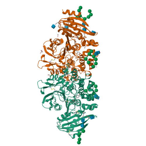
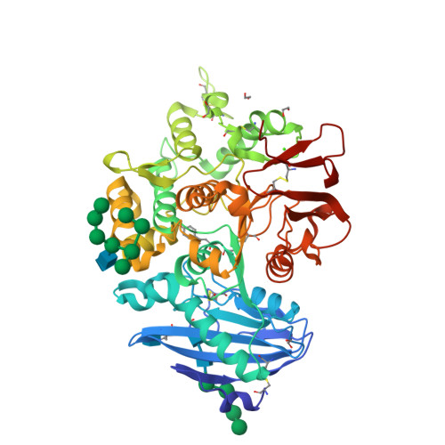
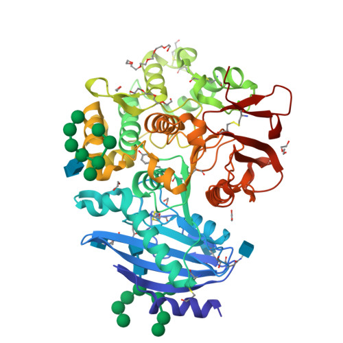
    Feruloyl esterases (FAEs) hydrolyze the ester bonds between hydroxycinnamic acids and arabinose residues of plant cell walls and exhibit considerable diversity in terms of substrate specificity. Here, we report the crystal structure of an FAE from Fusarium oxysporum (FoFaeC) at 1.7 Å resolution in complex with p-coumaric acid, which is the first ligand-bound structure of a tannase-like FAE. Our data reveal local conformational changes around the active site upon ligand binding, suggesting alternation between an active and a resting state of the enzyme. A swinging tyrosine residue appears to be gating the substrate binding pocket, while the lid domain of the protein exerts substrate specificity by means of a well-defined hydrophobic core that encases the phenyl moiety of the substrate.


  • Organizational Affiliation

    Industrial Biotechnology & Biocatalysis Group, Biotechnology Laboratory, School of Chemical Engineering, National Technical University of Athens, Greece.


Macromolecules
Find similar proteins by:  (by identity cutoff)  |  3D Structure
Entity ID: 1
MoleculeChains Sequence LengthOrganismDetailsImage
Carboxylic ester hydrolase
A, B
508Fusarium oxysporumMutation(s): 0 
Gene Names: faeC
EC: 3.1.1
UniProt
Find proteins for A0A2H3G048 (Fusarium oxysporum f. sp. radicis-cucumerinum)
Explore A0A2H3G048 
Go to UniProtKB:  A0A2H3G048
Entity Groups  
Sequence Clusters30% Identity50% Identity70% Identity90% Identity95% Identity100% Identity
UniProt GroupA0A2H3G048
Glycosylation
Glycosylation Sites: 3
Sequence Annotations
Expand
  • Reference Sequence
Oligosaccharides

Help

Entity ID: 2
MoleculeChains Length2D Diagram Glycosylation3D Interactions
alpha-D-mannopyranose-(1-2)-alpha-D-mannopyranose-(1-2)-alpha-D-mannopyranose-(1-3)-[alpha-D-mannopyranose-(1-2)-alpha-D-mannopyranose-(1-6)-[alpha-D-mannopyranose-(1-3)]alpha-D-mannopyranose-(1-6)]beta-D-mannopyranose-(1-4)-2-acetamido-2-deoxy-beta-D-glucopyranose-(1-4)-2-acetamido-2-deoxy-beta-D-glucopyranoseC,
D [auth G],
E [auth I]
10N-Glycosylation
Glycosylation Resources
GlyTouCan:  G40702WU
GlyCosmos:  G40702WU
GlyGen:  G40702WU
Entity ID: 3
MoleculeChains Length2D Diagram Glycosylation3D Interactions
alpha-D-mannopyranose-(1-2)-alpha-D-mannopyranose-(1-6)-alpha-D-mannopyranose-(1-6)-beta-D-mannopyranose-(1-4)-2-acetamido-2-deoxy-beta-D-glucopyranose-(1-4)-2-acetamido-2-deoxy-beta-D-glucopyranoseF [auth E]6N-Glycosylation
Glycosylation Resources
GlyTouCan:  G02763QD
GlyCosmos:  G02763QD
GlyGen:  G02763QD
Small Molecules
Ligands 7 Unique
IDChains Name / Formula / InChI Key2D Diagram3D Interactions
P6G
Query on P6G

Download Ideal Coordinates CCD File 
O [auth B]HEXAETHYLENE GLYCOL
C12 H26 O7
IIRDTKBZINWQAW-UHFFFAOYSA-N
NAG
Query on NAG

Download Ideal Coordinates CCD File 
H [auth A],
P [auth B],
X [auth B],
Y [auth B]
2-acetamido-2-deoxy-beta-D-glucopyranose
C8 H15 N O6
OVRNDRQMDRJTHS-FMDGEEDCSA-N
PG4
Query on PG4

Download Ideal Coordinates CCD File 
N [auth B]TETRAETHYLENE GLYCOL
C8 H18 O5
UWHCKJMYHZGTIT-UHFFFAOYSA-N
HC4 (Subject of Investigation/LOI)
Query on HC4

Download Ideal Coordinates CCD File 
G [auth A],
Q [auth B]
4'-HYDROXYCINNAMIC ACID
C9 H8 O3
NGSWKAQJJWESNS-ZZXKWVIFSA-N
PEG
Query on PEG

Download Ideal Coordinates CCD File 
V [auth B]DI(HYDROXYETHYL)ETHER
C4 H10 O3
MTHSVFCYNBDYFN-UHFFFAOYSA-N
EDO
Query on EDO

Download Ideal Coordinates CCD File 
I [auth A]
J [auth A]
K [auth A]
L [auth A]
R [auth B]
I [auth A],
J [auth A],
K [auth A],
L [auth A],
R [auth B],
S [auth B],
T [auth B],
U [auth B],
W [auth B],
Z [auth B]
1,2-ETHANEDIOL
C2 H6 O2
LYCAIKOWRPUZTN-UHFFFAOYSA-N
CA
Query on CA

Download Ideal Coordinates CCD File 
AA [auth B],
M [auth A]
CALCIUM ION
Ca
BHPQYMZQTOCNFJ-UHFFFAOYSA-N
Experimental Data & Validation

Experimental Data

  • Method: X-RAY DIFFRACTION
  • Resolution: 1.69 Å
  • R-Value Free: 0.217 
  • R-Value Work: 0.171 
  • R-Value Observed: 0.173 
  • Space Group: P 1 21 1
Unit Cell:
Length ( Å )Angle ( ˚ )
a = 67.704α = 90
b = 89.637β = 103.99
c = 116.978γ = 90
Software Package:
Software NamePurpose
REFMACrefinement
AutoProcessdata reduction
AutoProcessdata scaling
PHASERphasing

Structure Validation

View Full Validation Report


Ligand Structure Quality Assessment 


Entry History & Funding Information

Deposition Data


Funding OrganizationLocationGrant Number
Hellenic Foundation for Research and Innovation (HFRI)Greece03061
European CommissionEuropean UnioniNEXT discovery (PID 12346

Revision History  (Full details and data files)

  • Version 1.0: 2023-07-05
    Type: Initial release
  • Version 1.1: 2023-07-12
    Changes: Database references
  • Version 1.2: 2024-01-03
    Changes: Data collection, Database references