8BHA | pdb_00008bha

GABA-A receptor a5 homomer - a5V3 - Basmisanil - HR


Experimental Data Snapshot

  • Method: ELECTRON MICROSCOPY
  • Resolution: 2.67 Å
  • Aggregation State: PARTICLE 
  • Reconstruction Method: SINGLE PARTICLE 

wwPDB Validation 3D Report Full Report

Validation slider image for 8BHA

This is version 1.4 of the entry. See complete history

Literature

The molecular basis of drug selectivity for alpha 5 subunit-containing GABA A receptors.

Kasaragod, V.B.Malinauskas, T.Wahid, A.A.Lengyel, J.Knoflach, F.Hardwick, S.W.Jones, C.F.Chen, W.N.Lucas, X.El Omari, K.Chirgadze, D.Y.Aricescu, A.R.Cecere, G.Hernandez, M.C.Miller, P.S.

(2023) Nat Struct Mol Biol 30: 1936-1946

  • DOI: https://doi.org/10.1038/s41594-023-01133-1
  • Primary Citation Related Structures: 
    8BEJ, 8BGI, 8BHA, 8BHB, 8BHG, 8BHI, 8BHK, 8BHM, 8BHO, 8BHQ, 8BHR, 8BHS

  • PubMed Abstract: 

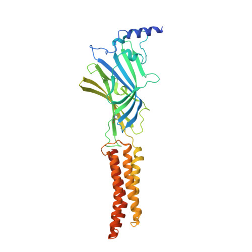
    α5 subunit-containing γ-aminobutyric acid type A (GABA A ) receptors represent a promising drug target for neurological and neuropsychiatric disorders. Altered expression and function contributes to neurodevelopmental disorders such as Dup15q and Angelman syndromes, developmental epilepsy and autism. Effective drug action without side effects is dependent on both α5-subtype selectivity and the strength of the positive or negative allosteric modulation (PAM or NAM). Here we solve structures of drugs bound to the α5 subunit. These define the molecular basis of binding and α5 selectivity of the β-carboline, methyl 6,7-dimethoxy-4-ethyl-β-carboline-3-carboxylate (DMCM), type II benzodiazepine NAMs, and a series of isoxazole NAMs and PAMs. For the isoxazole series, each molecule appears as an 'upper' and 'lower' moiety in the pocket. Structural data and radioligand binding data reveal a positional displacement of the upper moiety containing the isoxazole between the NAMs and PAMs. Using a hybrid molecule we directly measure the functional contribution of the upper moiety to NAM versus PAM activity. Overall, these structures provide a framework by which to understand distinct modulator binding modes and their basis of α5-subtype selectivity, appreciate structure-activity relationships, and empower future structure-based drug design campaigns.


  • Organizational Affiliation
    • Department of Pharmacology, University of Cambridge, Cambridge, UK.

Macromolecule Content 

  • Total Structure Weight: 208.62 kDa 
  • Atom Count: 13,805 
  • Modeled Residue Count: 1,680 
  • Deposited Residue Count: 1,800 
  • Unique protein chains: 1

Macromolecules

Find similar proteins by:|  3D Structure
Entity ID: 1
MoleculeChains  Sequence LengthOrganismDetailsImage
Gamma-aminobutyric acid receptor subunit alpha-5
A, B, C, D, E
360Homo sapiensMutation(s): 21 
Gene Names: GABRA5
Membrane Entity: Yes 
UniProt & NIH Common Fund Data Resources
Find proteins for P31644 (Homo sapiens)
Explore P31644 
Go to UniProtKB:  P31644
PHAROS:  P31644
GTEx:  ENSG00000186297 
Entity Groups
Sequence Clusters30% Identity50% Identity70% Identity90% Identity95% Identity100% Identity
UniProt GroupP31644
Glycosylation
Glycosylation Sites: 1Go to GlyGen: P31644-1
Sequence Annotations
Expand
Reference Sequence

Small Molecules

Ligands 2 Unique
IDChains Name / Formula / InChI Key2D Diagram3D Interactions
QI0
(Subject of Investigation/LOI)

Query on QI0


Download:Ideal Coordinates CCD File
G [auth A],
I [auth B],
K [auth C],
M [auth D],
O [auth E]
Basmisanil
C21 H20 F N3 O5 S
VCGRFBXVSFAGGA-UHFFFAOYSA-N
NAG

Query on NAG


Download:Ideal Coordinates CCD File
F [auth A],
H [auth B],
J [auth C],
L [auth D],
N [auth E]
2-acetamido-2-deoxy-beta-D-glucopyranose
C8 H15 N O6
OVRNDRQMDRJTHS-FMDGEEDCSA-N

Experimental Data & Validation

Experimental Data

  • Method: ELECTRON MICROSCOPY
  • Resolution: 2.67 Å
  • Aggregation State: PARTICLE 
  • Reconstruction Method: SINGLE PARTICLE 

Structure Validation

View Full Validation Report


Entry History 

& Funding Information

Deposition Data


Funding OrganizationLocationGrant Number
Other private--

Revision History  (Full details and data files)

  • Version 1.0: 2023-11-01
    Type: Initial release
  • Version 1.1: 2023-11-08
    Changes: Database references
  • Version 1.2: 2023-11-15
    Changes: Database references
  • Version 1.3: 2023-12-27
    Changes: Database references
  • Version 1.4: 2024-11-06
    Changes: Data collection, Structure summary