8BGL | pdb_00008bgl

Structure of the dimeric rsCherryRev1.4


Experimental Data Snapshot

  • Method: X-RAY DIFFRACTION
  • Resolution: 2.00 Å
  • R-Value Free: 
    0.231 (Depositor), 0.232 (DCC) 
  • R-Value Work: 
    0.188 (Depositor), 0.188 (DCC) 
  • R-Value Observed: 
    0.190 (Depositor) 

Starting Model: experimental
View more details

wwPDB Validation 3D Report Full Report

Validation slider image for 8BGL

This is version 3.1 of the entry. See complete history

Literature

An unusual disulfide-linked dimerization in the fluorescent protein rsCherryRev1.4.

Bui, T.Y.H.Dedecker, P.Van Meervelt, L.

(2023) Acta Crystallogr F Struct Biol Commun 79: 38-44

  • DOI: https://doi.org/10.1107/S2053230X23000572
  • Primary Citation Related Structures: 
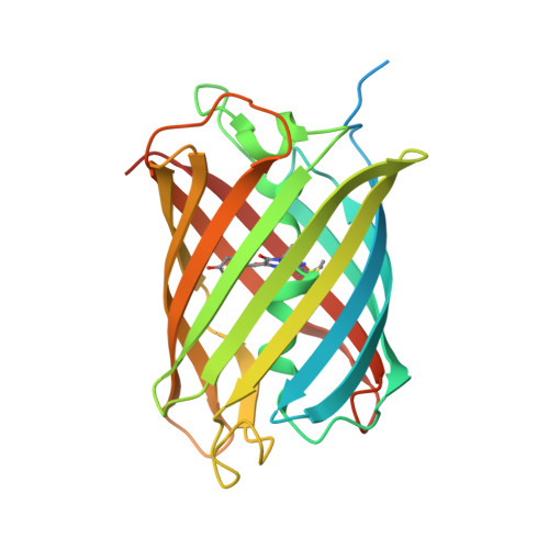
    8BGL

  • PubMed Abstract: 

    rsCherryRev1.4 has been reported as one of the reversibly photoswitchable variants of mCherry, and is an improved version with a faster off-switching speed and lower switching fatigue at high light intensities than its precursor rsCherryRev. However, rsCherryRev1.4 still has some limitations such as a tendency to dimerize as well as complex photophysical properties. Here, the crystal structure of rsCherryRev1.4 was determined at a resolution of 2 Å and it was discovered that it forms a dimer that shows disulfide bonding between the protomers. Mutagenesis, gel electrophoresis and size-exclusion chromatography strongly implicate Cys24 in this process. Replacing Cys24 in rsCherryRev1.4 resulted in a much lower tendency towards dimerization, while introducing Cys24 into mCherry correspondingly increased its dimerization. In principle, this finding opens the possibility of developing redox sensors based on controlled dimerization via disulfide cross-linking in fluorescent proteins, even though the actual application of engineering such sensors still requires additional research.


  • Organizational Affiliation
    • Biochemistry, Molecular and Structural Biology, Department of Chemistry, KU Leuven, 3001 Leuven, Belgium.

Macromolecule Content 

  • Total Structure Weight: 61.6 kDa 
  • Atom Count: 3,792 
  • Modeled Residue Count: 436 
  • Deposited Residue Count: 538 
  • Unique protein chains: 1

Macromolecules

Find similar proteins by:|  3D Structure
Entity ID: 1
MoleculeChains  Sequence LengthOrganismDetailsImage
PAmCherry1 protein
A, B
267Discosoma sp.Mutation(s): 2 
Gene Names: PAmCherryPAmCherry1
UniProt
Find proteins for Q9U6Y8 (Discosoma sp.)
Explore Q9U6Y8 
Go to UniProtKB:  Q9U6Y8
Entity Groups
Sequence Clusters30% Identity50% Identity70% Identity90% Identity95% Identity100% Identity
UniProt GroupQ9U6Y8
Sequence Annotations
Expand
Reference Sequence

Small Molecules

Experimental Data & Validation

Experimental Data

  • Method: X-RAY DIFFRACTION
  • Resolution: 2.00 Å
  • R-Value Free:  0.231 (Depositor), 0.232 (DCC) 
  • R-Value Work:  0.188 (Depositor), 0.188 (DCC) 
  • R-Value Observed: 0.190 (Depositor) 
Space Group: I 1 2 1
Unit Cell:
Length ( Å )Angle ( ˚ )
a = 98.104α = 90
b = 48.822β = 101.16
c = 99.12γ = 90
Software Package:
Software NamePurpose
PHENIXrefinement
XDSdata reduction
Aimlessdata scaling
PDB_EXTRACTdata extraction
PHASERphasing

Structure Validation

View Full Validation Report


Entry History 

& Funding Information

Deposition Data


Funding OrganizationLocationGrant Number
Other government911 project
KU LeuvenBelgium--

Revision History  (Full details and data files)

  • Version 1.0: 2023-02-15
    Type: Initial release
  • Version 1.1: 2023-04-05
    Changes: Database references
  • Version 2.0: 2023-11-15
    Changes: Atomic model, Data collection, Derived calculations
  • Version 2.1: 2024-02-07
    Changes: Refinement description
  • Version 3.0: 2024-07-24
    Changes: Atomic model, Data collection, Database references, Derived calculations, Polymer sequence, Refinement description, Source and taxonomy, Structure summary
  • Version 3.1: 2024-10-16
    Changes: Structure summary