8BCS | pdb_00008bcs

X-ray crystal structure of a de novo designed helix-loop-helix homodimer in an anti arrangement, CC-HP1.0


Experimental Data Snapshot

  • Method: X-RAY DIFFRACTION
  • Resolution: 2.10 Å
  • R-Value Free: 
    0.265 (Depositor), 0.267 (DCC) 
  • R-Value Work: 
    0.237 (Depositor), 0.239 (DCC) 
  • R-Value Observed: 
    0.240 (Depositor) 

Starting Model: experimental
View more details

wwPDB Validation 3D Report Full Report

Validation slider image for 8BCS

This is version 1.3 of the entry. See complete history

Literature

Design and Selection of Heterodimerizing Helical Hairpins for Synthetic Biology.

Smith, A.J.Naudin, E.A.Edgell, C.L.Baker, E.G.Mylemans, B.FitzPatrick, L.Herman, A.Rice, H.M.Andrews, D.M.Tigue, N.Woolfson, D.N.Savery, N.J.

(2023) ACS Synth Biol 12: 1845-1858

  • DOI: https://doi.org/10.1021/acssynbio.3c00231
  • Primary Citation Related Structures: 
    8BCS, 8BCT

  • PubMed Abstract: 

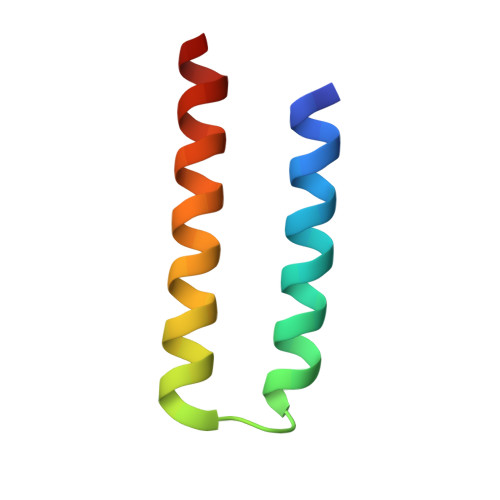
    Synthetic biology applications would benefit from protein modules of reduced complexity that function orthogonally to cellular components. As many subcellular processes depend on peptide-protein or protein-protein interactions, de novo designed polypeptides that can bring together other proteins controllably are particularly useful. Thanks to established sequence-to-structure relationships, helical bundles provide good starting points for such designs. Typically, however, such designs are tested in vitro and function in cells is not guaranteed. Here, we describe the design, characterization, and application of de novo helical hairpins that heterodimerize to form 4-helix bundles in cells. Starting from a rationally designed homodimer, we construct a library of helical hairpins and identify complementary pairs using bimolecular fluorescence complementation in E. coli . We characterize some of the pairs using biophysics and X-ray crystallography to confirm heterodimeric 4-helix bundles. Finally, we demonstrate the function of an exemplar pair in regulating transcription in both E. coli and mammalian cells.


  • Organizational Affiliation
    • School of Biochemistry, University of Bristol, Bristol BS8 1TD, U.K.

Macromolecule Content 

  • Total Structure Weight: 5.33 kDa 
  • Atom Count: 330 
  • Modeled Residue Count: 49 
  • Deposited Residue Count: 51 
  • Unique protein chains: 1

Macromolecules

Find similar proteins by:|  3D Structure
Entity ID: 1
MoleculeChains  Sequence LengthOrganismDetailsImage
CC-HP1.051synthetic constructMutation(s): 0 

Small Molecules

Ligands 1 Unique
IDChains Name / Formula / InChI Key2D Diagram3D Interactions
ACT

Query on ACT


Download:Ideal Coordinates CCD File
B [auth A]ACETATE ION
C2 H3 O2
QTBSBXVTEAMEQO-UHFFFAOYSA-M

Experimental Data & Validation

Experimental Data

  • Method: X-RAY DIFFRACTION
  • Resolution: 2.10 Å
  • R-Value Free:  0.265 (Depositor), 0.267 (DCC) 
  • R-Value Work:  0.237 (Depositor), 0.239 (DCC) 
  • R-Value Observed: 0.240 (Depositor) 
Space Group: P 31 2 1
Unit Cell:
Length ( Å )Angle ( ˚ )
a = 36.438α = 90
b = 36.438β = 90
c = 59.329γ = 120
Software Package:
Software NamePurpose
PHENIXrefinement
DIALSdata reduction
Aimlessdata scaling
PHASERphasing

Structure Validation

View Full Validation Report


Entry History 

& Funding Information

Deposition Data


Funding OrganizationLocationGrant Number
Biotechnology and Biological Sciences Research Council (BBSRC)United KingdomBB/S002820/1
Biotechnology and Biological Sciences Research Council (BBSRC)United KingdomBB/V006231/1
Max Planck Bristol Centre for Minimal Biology - University of BristolUnited Kingdom--

Revision History  (Full details and data files)

  • Version 1.0: 2023-06-07
    Type: Initial release
  • Version 1.1: 2023-06-28
    Changes: Database references
  • Version 1.2: 2024-02-07
    Changes: Data collection, Refinement description
  • Version 1.3: 2024-10-16
    Changes: Structure summary