8BBQ | pdb_00008bbq

Determination of the structure of active tyrosinase from bacterium Verrucomicrobium spinosum


Experimental Data Snapshot

  • Method: X-RAY DIFFRACTION
  • Resolution: 1.43 Å
  • R-Value Free: 
    0.200 (Depositor), 0.203 (DCC) 
  • R-Value Work: 
    0.169 (Depositor), 0.177 (DCC) 
  • R-Value Observed: 
    0.171 (Depositor) 

Starting Models: experimental
View more details

wwPDB Validation 3D Report Full Report

Validation slider image for 8BBQ

This is version 1.2 of the entry. See complete history

Literature

The Crystal Structure of Tyrosinase from Verrucomicrobium spinosum Reveals It to Be an Atypical Bacterial Tyrosinase.

Fekry, M.Dave, K.K.Badgujar, D.Hamnevik, E.Aurelius, O.Dobritzsch, D.Danielson, U.H.

(2023) Biomolecules 13

  • DOI: https://doi.org/10.3390/biom13091360
  • Primary Citation Related Structures: 
    8BBQ, 8BBR

  • PubMed Abstract: 

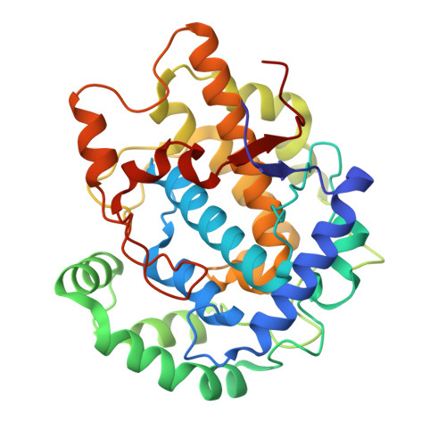
    Tyrosinases belong to the type-III copper enzyme family, which is involved in melanin production in a wide range of organisms. Despite similar overall characteristics and functions, their structures, activities, substrate specificities and regulation vary. The tyrosinase from the bacterium Verrucomicrobium spinosum ( vs Tyr) is produced as a pre-pro-enzyme in which a C-terminal extension serves as an inactivation domain. It does not require a caddie protein for copper ion incorporation, which makes it similar to eukaryotic tyrosinases. To gain an understanding of the catalytic machinery and regulation of vs Tyr activity, we determined the structure of the catalytically active "core domain" of vs Tyr by X-ray crystallography. The analysis showed that vs Tyr is an atypical bacterial tyrosinase not only because it is independent of a caddie protein but also because it shows the highest structural (and sequence) similarity to plant-derived members of the type-III copper enzyme family and is more closely related to fungal tyrosinases regarding active site features. By modelling the structure of the pre-pro-enzyme using AlphaFold, we observed that Phe453, located in the C-terminal extension, is appropriately positioned to function as a "gatekeeper" residue. Our findings raise questions concerning the evolutionary origin of vs Tyr.


  • Organizational Affiliation
    • Department of Chemistry-BMC, Uppsala University, SE 751 23 Uppsala, Sweden.

Macromolecule Content 

  • Total Structure Weight: 74.61 kDa 
  • Atom Count: 5,787 
  • Modeled Residue Count: 645 
  • Deposited Residue Count: 648 
  • Unique protein chains: 1

Macromolecules

Find similar proteins by:|  3D Structure
Entity ID: 1
MoleculeChains  Sequence LengthOrganismDetailsImage
Core tyrosinase
A, B
324Verrucomicrobium spinosumMutation(s): 0 
EC: 1.14.18.1
UniProt
Find proteins for A0AAJ6N653 (Verrucomicrobium spinosum)
Explore A0AAJ6N653 
Go to UniProtKB:  A0AAJ6N653
Entity Groups
Sequence Clusters30% Identity50% Identity70% Identity90% Identity95% Identity100% Identity
UniProt GroupA0AAJ6N653
Sequence Annotations
Expand
Reference Sequence

Small Molecules

Ligands 3 Unique
IDChains Name / Formula / InChI Key2D Diagram3D Interactions
SO4

Query on SO4


Download:Ideal Coordinates CCD File
G [auth A],
M [auth B]
SULFATE ION
O4 S
QAOWNCQODCNURD-UHFFFAOYSA-L
GOL

Query on GOL


Download:Ideal Coordinates CCD File
C [auth A]
D [auth A]
E [auth A]
F [auth A]
J [auth B]
C [auth A],
D [auth A],
E [auth A],
F [auth A],
J [auth B],
K [auth B],
L [auth B]
GLYCEROL
C3 H8 O3
PEDCQBHIVMGVHV-UHFFFAOYSA-N
CU
(Subject of Investigation/LOI)

Query on CU


Download:Ideal Coordinates CCD File
H [auth A],
I [auth A],
N [auth B],
O [auth B]
COPPER (II) ION
Cu
JPVYNHNXODAKFH-UHFFFAOYSA-N

Experimental Data & Validation

Experimental Data

  • Method: X-RAY DIFFRACTION
  • Resolution: 1.43 Å
  • R-Value Free:  0.200 (Depositor), 0.203 (DCC) 
  • R-Value Work:  0.169 (Depositor), 0.177 (DCC) 
  • R-Value Observed: 0.171 (Depositor) 
Space Group: C 1 2 1
Unit Cell:
Length ( Å )Angle ( ˚ )
a = 84.813α = 90
b = 63.422β = 96.83
c = 116.02γ = 90
Software Package:
Software NamePurpose
HKL-2000data reduction
Aimlessdata scaling
PHENIXrefinement
PDB_EXTRACTdata extraction
MOLREPphasing

Structure Validation

View Full Validation Report


Entry History 

& Funding Information

Deposition Data


Funding OrganizationLocationGrant Number
Other privateChina--

Revision History  (Full details and data files)

  • Version 1.0: 2023-09-20
    Type: Initial release
  • Version 1.1: 2023-10-11
    Changes: Database references
  • Version 1.2: 2026-03-04
    Changes: Refinement description, Structure summary