8BAR | pdb_00008bar

E. coli C7 DarT1 in complex with ADP-ribosylated ssDNA and nicotinamide


Experimental Data Snapshot

  • Method: X-RAY DIFFRACTION
  • Resolution: 1.63 Å
  • R-Value Free: 
    0.178 (Depositor), 0.187 (DCC) 
  • R-Value Work: 
    0.155 (Depositor), 0.166 (DCC) 

Starting Model: experimental
View more details

wwPDB Validation 3D Report Full Report

Validation slider image for 8BAR

Ligand Structure Quality Assessment 


This is version 1.2 of the entry. See complete history

Literature

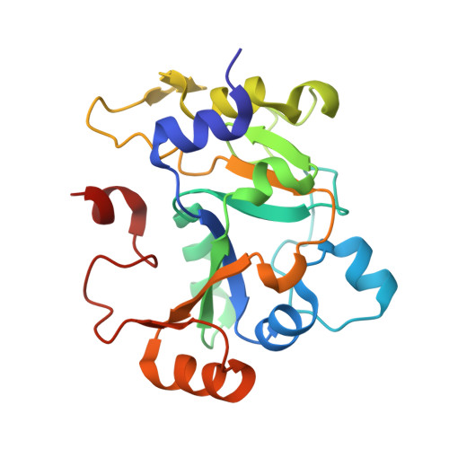
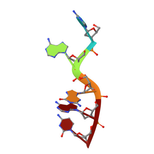
Molecular basis for the reversible ADP-ribosylation of guanosine bases.

Schuller, M.Raggiaschi, R.Mikolcevic, P.Rack, J.G.M.Ariza, A.Zhang, Y.Ledermann, R.Tang, C.Mikoc, A.Ahel, I.

(2023) Mol Cell 83: 2303

  • DOI: https://doi.org/10.1016/j.molcel.2023.06.013
  • Primary Citation Related Structures: 
    8BAQ, 8BAR, 8BAS, 8BAT, 8BAU

  • PubMed Abstract: 

    Modification of nucleic acids by ADP-ribosylation is catalyzed by various ADP-ribosyltransferases, including the DarT enzyme. The latter is part of the bacterial toxin-antitoxin (TA) system DarTG, which was shown to provide control of DNA replication and bacterial growth as well as protection against bacteriophages. Two subfamilies have been identified, DarTG1 and DarTG2, which are distinguished by their associated antitoxins. While DarTG2 catalyzes reversible ADP-ribosylation of thymidine bases employing a macrodomain as antitoxin, the DNA ADP-ribosylation activity of DarTG1 and the biochemical function of its antitoxin, a NADAR domain, are as yet unknown. Using structural and biochemical approaches, we show that DarT1-NADAR is a TA system for reversible ADP-ribosylation of guanosine bases. DarT1 evolved the ability to link ADP-ribose to the guanine amino group, which is specifically hydrolyzed by NADAR. We show that guanine de-ADP-ribosylation is also conserved among eukaryotic and non-DarT-associated NADAR members, indicating a wide distribution of reversible guanine modifications beyond DarTG systems.


  • Organizational Affiliation
    • Sir William Dunn School of Pathology, University of Oxford, Oxford, UK.

Macromolecule Content 

  • Total Structure Weight: 26.81 kDa 
  • Atom Count: 2,250 
  • Modeled Residue Count: 213 
  • Deposited Residue Count: 213 
  • Unique protein chains: 1
  • Unique nucleic acid chains: 1

Macromolecules


Find similar proteins by:|  3D Structure
Entity ID: 1
MoleculeChains  Sequence LengthOrganismDetailsImage
E. coli C7 DarT1208Escherichia coliMutation(s): 0 
Gene Names: C3F40_23400
UniProt
Find proteins for A0A794RBZ4 (Escherichia coli)
Explore A0A794RBZ4 
Go to UniProtKB:  A0A794RBZ4
Entity Groups
Sequence Clusters30% Identity50% Identity70% Identity90% Identity95% Identity100% Identity
UniProt GroupA0A794RBZ4
Sequence Annotations
Expand
Reference Sequence
Find similar nucleic acids by:  Sequence
Entity ID: 2
MoleculeChains LengthOrganismImage
DNA (5'-D(*AP*AP*GP*AP*C)-3')B [auth D]5Escherichia coli
Sequence Annotations
Expand
Reference Sequence

Small Molecules

Ligands 3 Unique
IDChains Name / Formula / InChI Key2D Diagram3D Interactions
APR
(Subject of Investigation/LOI)

Query on APR


Download:Ideal Coordinates CCD File
K [auth D]ADENOSINE-5-DIPHOSPHORIBOSE
C15 H23 N5 O14 P2
SRNWOUGRCWSEMX-KEOHHSTQSA-N
NCA

Query on NCA


Download:Ideal Coordinates CCD File
J [auth A]NICOTINAMIDE
C6 H6 N2 O
DFPAKSUCGFBDDF-UHFFFAOYSA-N
EDO

Query on EDO


Download:Ideal Coordinates CCD File
C [auth A]
D [auth A]
E [auth A]
F [auth A]
G [auth A]
C [auth A],
D [auth A],
E [auth A],
F [auth A],
G [auth A],
H [auth A],
I [auth A]
1,2-ETHANEDIOL
C2 H6 O2
LYCAIKOWRPUZTN-UHFFFAOYSA-N

Experimental Data & Validation

Experimental Data

  • Method: X-RAY DIFFRACTION
  • Resolution: 1.63 Å
  • R-Value Free:  0.178 (Depositor), 0.187 (DCC) 
  • R-Value Work:  0.155 (Depositor), 0.166 (DCC) 
Space Group: P 43 21 2
Unit Cell:
Length ( Å )Angle ( ˚ )
a = 61.759α = 90
b = 61.759β = 90
c = 215.039γ = 90
Software Package:
Software NamePurpose
REFMACrefinement
DIALSdata reduction
Aimlessdata scaling
PHASERphasing

Structure Validation

View Full Validation Report


Ligand Structure Quality Assessment 


Entry History 

& Funding Information

Deposition Data


Funding OrganizationLocationGrant Number
Biotechnology and Biological Sciences Research Council (BBSRC)United KingdomBB/R007195/1

Revision History  (Full details and data files)

  • Version 1.0: 2023-07-12
    Type: Initial release
  • Version 1.1: 2023-07-19
    Changes: Data collection, Database references
  • Version 1.2: 2024-02-07
    Changes: Data collection, Refinement description