8BA8 | pdb_00008ba8

CryoEM structure of GroEL-ADP.BeF3-Rubisco.


Experimental Data Snapshot

  • Method: ELECTRON MICROSCOPY
  • Resolution: 3.40 Å
  • Aggregation State: PARTICLE 
  • Reconstruction Method: SINGLE PARTICLE 

wwPDB Validation 3D Report Full Report

Validation slider image for 8BA8

This is version 1.1 of the entry. See complete history

Literature

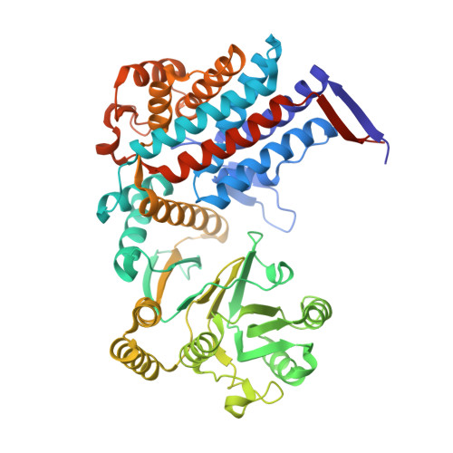
Structural basis of substrate progression through the bacterial chaperonin cycle.

Gardner, S.Darrow, M.C.Lukoyanova, N.Thalassinos, K.Saibil, H.R.

(2023) Proc Natl Acad Sci U S A 120: e2308933120-e2308933120

  • DOI: https://doi.org/10.1073/pnas.2308933120
  • Primary Citation Related Structures: 
    8BA7, 8BA8, 8BA9, 8BAA

  • PubMed Abstract: 

    The bacterial chaperonin GroEL-GroES promotes protein folding through ATP-regulated cycles of substrate protein binding, encapsulation, and release. Here, we have used cryoEM to determine structures of GroEL, GroEL-ADP·BeF 3 , and GroEL-ADP·AlF 3 -GroES all complexed with the model substrate Rubisco. Our structures provide a series of snapshots that show how the conformation and interactions of non-native Rubisco change as it proceeds through the GroEL-GroES reaction cycle. We observe specific charged and hydrophobic GroEL residues forming strong initial contacts with non-native Rubisco. Binding of ATP or ADP·BeF 3 to GroEL-Rubisco results in the formation of an intermediate GroEL complex displaying striking asymmetry in the ATP/ADP·BeF 3 -bound ring. In this ring, four GroEL subunits bind Rubisco and the other three are in the GroES-accepting conformation, suggesting how GroEL can recruit GroES without releasing bound substrate. Our cryoEM structures of stalled GroEL-ADP·AlF 3 -Rubisco-GroES complexes show Rubisco folding intermediates interacting with GroEL-GroES via different sets of residues.


  • Organizational Affiliation
    • Institute of Structural and Molecular Biology, Department of Biological Sciences, Birkbeck, University of London, London WC1E 7HX, United Kingdom.

Macromolecule Content 

  • Total Structure Weight: 810.81 kDa 
  • Atom Count: 54,425 
  • Modeled Residue Count: 7,336 
  • Deposited Residue Count: 7,672 
  • Unique protein chains: 1

Macromolecules

Find similar proteins by:|  3D Structure
Entity ID: 1
MoleculeChains  Sequence LengthOrganismDetailsImage
Chaperonin GroEL
A, B, C, D, E
A, B, C, D, E, F, G, H, I, J, K, L, M, N
548Escherichia coli K-12Mutation(s): 0 
Gene Names: groELgroLmopAb4143JW4103
EC: 5.6.1.7
UniProt
Find proteins for P0A6F5 (Escherichia coli (strain K12))
Explore P0A6F5 
Go to UniProtKB:  P0A6F5
Entity Groups
Sequence Clusters30% Identity50% Identity70% Identity90% Identity95% Identity100% Identity
UniProt GroupP0A6F5
Sequence Annotations
Expand
Reference Sequence

Small Molecules

Ligands 4 Unique
IDChains Name / Formula / InChI Key2D Diagram3D Interactions
ADP

Query on ADP


Download:Ideal Coordinates CCD File
AB [auth K]
CB [auth L]
DA [auth D]
HA [auth E]
HB [auth M]
AB [auth K],
CB [auth L],
DA [auth D],
HA [auth E],
HB [auth M],
IB [auth N],
JA [auth F],
MA [auth G],
P [auth A],
SA [auth H],
TA [auth I],
V [auth B],
W [auth C],
WA [auth J]
ADENOSINE-5'-DIPHOSPHATE
C10 H15 N5 O10 P2
XTWYTFMLZFPYCI-KQYNXXCUSA-N
BEF

Query on BEF


Download:Ideal Coordinates CCD File
AA [auth D]
EA [auth E]
IA [auth F]
O [auth A]
OA [auth G]
AA [auth D],
EA [auth E],
IA [auth F],
O [auth A],
OA [auth G],
U [auth B],
Y [auth C]
BERYLLIUM TRIFLUORIDE ION
Be F3
OGIAHMCCNXDTIE-UHFFFAOYSA-K
K

Query on K


Download:Ideal Coordinates CCD File
BB [auth K]
CA [auth D]
DB [auth L]
FA [auth E]
GB [auth M]
BB [auth K],
CA [auth D],
DB [auth L],
FA [auth E],
GB [auth M],
JB [auth N],
KA [auth F],
PA [auth G],
QA [auth H],
R [auth A],
T [auth B],
VA [auth I],
YA [auth J],
Z [auth C]
POTASSIUM ION
K
NPYPAHLBTDXSSS-UHFFFAOYSA-N
MG

Query on MG


Download:Ideal Coordinates CCD File
BA [auth D]
EB [auth L]
FB [auth M]
GA [auth E]
KB [auth N]
BA [auth D],
EB [auth L],
FB [auth M],
GA [auth E],
KB [auth N],
LA [auth F],
NA [auth G],
Q [auth A],
RA [auth H],
S [auth B],
UA [auth I],
X [auth C],
XA [auth J],
ZA [auth K]
MAGNESIUM ION
Mg
JLVVSXFLKOJNIY-UHFFFAOYSA-N

Experimental Data & Validation

Experimental Data

  • Method: ELECTRON MICROSCOPY
  • Resolution: 3.40 Å
  • Aggregation State: PARTICLE 
  • Reconstruction Method: SINGLE PARTICLE 
EM Software:
TaskSoftware PackageVersion
RECONSTRUCTIONRELION3.1

Structure Validation

View Full Validation Report


Entry History 

& Funding Information

Deposition Data


Funding OrganizationLocationGrant Number
Biotechnology and Biological Sciences Research Council (BBSRC)United KingdomBB/M009513/1

Revision History  (Full details and data files)

  • Version 1.0: 2023-12-27
    Type: Initial release
  • Version 1.1: 2026-02-25
    Changes: Data collection, Database references, Structure summary