DNA-binding and protein structure of nuclear factors likely acting in genetic information processing in the Paulinella chromatophore.
Macorano, L., Binny, T.M., Spiegl, T., Klimenko, V., Singer, A., Oberleitner, L., Applegate, V., Seyffert, S., Stefanski, A., Gremer, L., Gertzen, C.G.W., Hoppner, A., Smits, S.H.J., Nowack, E.C.M.(2023) Proc Natl Acad Sci U S A 120: e2221595120-e2221595120
- PubMed: 37364116 Search on PubMedSearch on PubMed Central
- DOI: https://doi.org/10.1073/pnas.2221595120
- Primary Citation Related Structures:
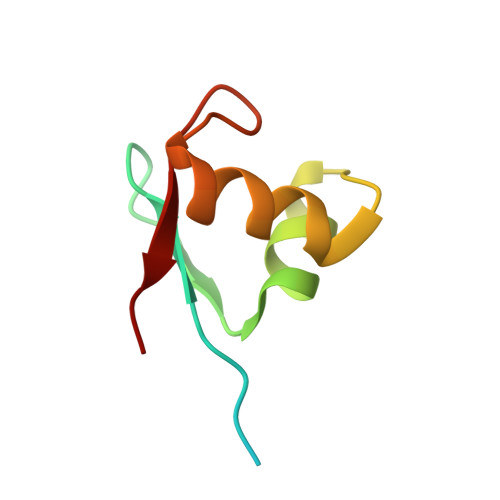
8B6E - PubMed Abstract:
The chromatophores in Paulinella are evolutionary-early-stage photosynthetic organelles. Biological processes in chromatophores depend on a combination of chromatophore and nucleus-encoded proteins. Interestingly, besides proteins carrying chromatophore-targeting signals, a large arsenal of short chromatophore-targeted proteins (sCTPs; <90 amino acids) without recognizable targeting signals were found in chromatophores. This situation resembles endosymbionts in plants and insects that are manipulated by host-derived antimicrobial peptides. Previously, we identified an expanded family of sCTPs of unknown function, named here "DNA-binding (DB)-sCTPs". DB-sCTPs contain a ~45 amino acid motif that is conserved in some bacterial proteins with predicted functions in DNA processing. Here, we explored antimicrobial activity, DNA-binding capacity, and structures of three purified recombinant DB-sCTPs. All three proteins exhibited antimicrobial activity against bacteria involving membrane permeabilization, and bound to bacterial lipids in vitro. A combination of in vitro assays demonstrated binding of recombinant DB-sCTPs to chromatophore-derived genomic DNA sequences with an affinity in the low nM range. Additionally, we report the 1.2 Å crystal structure of one DB-sCTP. In silico docking studies suggest that helix α2 inserts into the DNA major grove and the exposed residues, that are highly variable between different DB-sCTPs, confer interaction with the DNA bases. Identification of photosystem II subunit CP43 as a potential interaction partner of one DB-sCTP, suggests DB-sCTPs to be involved in more complex regulatory mechanisms. We hypothesize that membrane binding of DB-sCTPs is related to their import into chromatophores. Once inside, they interact with the chromatophore genome potentially providing nuclear control over genetic information processing.
- Institute of Microbial Cell Biology, Department of Biology, Heinrich Heine University Düsseldorf, 40225 Düsseldorf, Germany.
Organizational Affiliation: