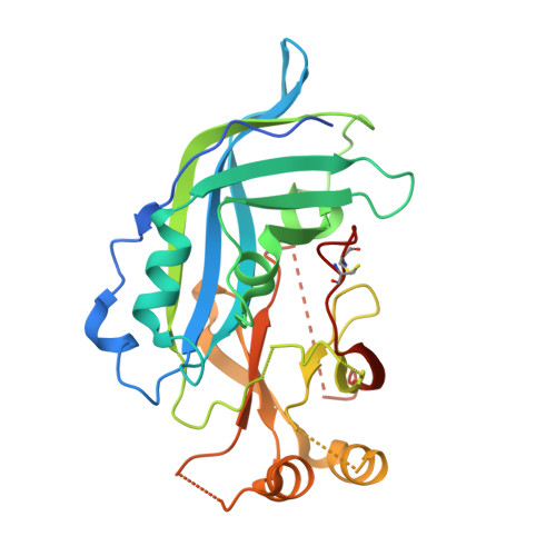
Development of a Crystallographic Screening to Identify Sudan Virus VP40 Ligands.
Werner, A.D., Krapoth, N., Norris, M.J., Heine, A., Klebe, G., Saphire, E.O., Becker, S.(2024) ACS Omega 9: 33193-33203
- PubMed: 39100314
- DOI: https://doi.org/10.1021/acsomega.4c04829
- Primary Citation of Related Structures:
8B1S, 8B2U - PubMed Abstract:
The matrix protein VP40 of the highly pathogenic Sudan virus (genus Orthoebolavirus ) is a multifunctional protein responsible for the recruitment of viral nucleocapsids to the plasma membrane and the budding of infectious virions. In addition to its role in assembly, VP40 also downregulates viral genome replication and transcription. VP40's existence in various homo-oligomeric states is presumed to underpin its diverse functional capabilities during the viral life cycle. Given the absence of licensed therapeutics targeting the Sudan virus, our study focused on inhibiting VP40 dimers, the structural precursors to critical higher-order oligomers, as a novel antiviral strategy. We have established a crystallographic screening pipeline for the identification of small-molecule fragments capable of binding to VP40. Dimeric VP40 of the Sudan virus was recombinantly expressed in bacteria, purified, crystallized, and soaked in a solution of 96 different preselected fragments. Salicylic acid was identified as a crystallographic hit with clear electron density in the pocket between the N- and the C-termini of the VP40 dimer. The binding interaction is predominantly coordinated by amino acid residues leucine 158 (L158) and arginine 214 (R214), which are key in stabilizing salicylic acid within the binding pocket. While salicylic acid displayed minimal impact on the functional aspects of VP40, we delved deeper into characterizing the druggability of the identified binding pocket. We analyzed the influence of residues L158 and R214 on the formation of virus-like particles and viral RNA synthesis. Site-directed mutagenesis of these residues to alanine markedly affected both VP40's budding activity and its effect on viral RNA synthesis, underscoring the potential of the salicylic acid binding pocket as a drug target. In summary, our findings lay the foundation for structure-guided drug design to provide lead compounds against Sudan virus VP40.
- Institute for Virology, University of Marburg, D-35043 Marburg, Hessen, Germany.
Organizational Affiliation:


