8B29 | pdb_00008b29

Human carbonic anhydrase II containing 6-fluorotryptophanes.


Experimental Data Snapshot

  • Method: X-RAY DIFFRACTION
  • Resolution: 1.70 Å
  • R-Value Free: 
    0.210 (Depositor), 0.210 (DCC) 
  • R-Value Work: 
    0.162 (Depositor), 0.162 (DCC) 
  • R-Value Observed: 
    0.165 (Depositor) 

Starting Model: experimental
View more details

wwPDB Validation 3D Report Full Report

Validation slider image for 8B29

This is version 1.4 of the entry. See complete history

Literature

Direct Expression of Fluorinated Proteins in Human Cells for 19 F In-Cell NMR Spectroscopy.

Pham, L.B.T.Costantino, A.Barbieri, L.Calderone, V.Luchinat, E.Banci, L.

(2023) J Am Chem Soc 145: 1389-1399

  • DOI: https://doi.org/10.1021/jacs.2c12086
  • Primary Citation Related Structures: 
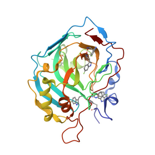
    8B29

  • PubMed Abstract: 

    In-cell NMR spectroscopy is a powerful approach to study protein structure and function in the native cellular environment. It provides precious insights into the folding, maturation, interactions, and ligand binding of important pharmacological targets directly in human cells. However, its widespread application is hampered by the fact that soluble globular proteins often interact with large cellular components, causing severe line broadening in conventional heteronuclear NMR experiments. 19 F NMR can overcome this issue, as fluorine atoms incorporated in proteins can be detected by simple background-free 1D NMR spectra. Here, we show that fluorinated amino acids can be easily incorporated in proteins expressed in human cells by employing a medium switch strategy. This straightforward approach allows the incorporation of different fluorinated amino acids in the protein of interest, reaching fluorination efficiencies up to 60%, as confirmed by mass spectrometry and X-ray crystallography. The versatility of the approach is shown by performing 19 F in-cell NMR on several proteins, including those that would otherwise be invisible by 1 H- 15 N in-cell NMR. We apply the approach to observe the interaction between an intracellular target, carbonic anhydrase 2, and its inhibitors, and to investigate how the formation of a complex between superoxide dismutase 1 and its chaperone CCS modulates the interaction of the chaperone subunit with the cellular environment.


  • Organizational Affiliation
    • CERM─Magnetic Resonance Center, Università degli Studi di Firenze, Via Luigi Sacconi 6, 50019Sesto Fiorentino, Italy.

Macromolecule Content 

  • Total Structure Weight: 29.26 kDa 
  • Atom Count: 2,354 
  • Modeled Residue Count: 258 
  • Deposited Residue Count: 258 
  • Unique protein chains: 1

Macromolecules

Find similar proteins by:|  3D Structure
Entity ID: 1
MoleculeChains  Sequence LengthOrganismDetailsImage
Carbonic anhydrase 2258Homo sapiensMutation(s): 0 
Gene Names: CA2
EC: 4.2.1.1 (PDB Primary Data), 4.2.1.69 (PDB Primary Data)
UniProt & NIH Common Fund Data Resources
Find proteins for P00918 (Homo sapiens)
Explore P00918 
Go to UniProtKB:  P00918
PHAROS:  P00918
GTEx:  ENSG00000104267 
Entity Groups
Sequence Clusters30% Identity50% Identity70% Identity90% Identity95% Identity100% Identity
UniProt GroupP00918
Sequence Annotations
Expand
Reference Sequence

Small Molecules

Modified Residues  1 Unique
IDChains TypeFormula2D DiagramParent
FT6
Query on FT6
A
L-PEPTIDE LINKINGC11 H11 F N2 O2TRP

Experimental Data & Validation

Experimental Data

  • Method: X-RAY DIFFRACTION
  • Resolution: 1.70 Å
  • R-Value Free:  0.210 (Depositor), 0.210 (DCC) 
  • R-Value Work:  0.162 (Depositor), 0.162 (DCC) 
  • R-Value Observed: 0.165 (Depositor) 
Space Group: P 1 21 1
Unit Cell:
Length ( Å )Angle ( ˚ )
a = 42.2α = 90
b = 41.22β = 104.33
c = 71.96γ = 90
Software Package:
Software NamePurpose
PHENIXrefinement
PDB_EXTRACTdata extraction
XDSdata reduction
XSCALEdata scaling
MOLREPphasing

Structure Validation

View Full Validation Report


Entry History 

& Funding Information

Deposition Data


Funding OrganizationLocationGrant Number
Ministero dell Universita e della RicercaItaly--

Revision History  (Full details and data files)

  • Version 1.0: 2023-01-18
    Type: Initial release
  • Version 1.1: 2023-01-25
    Changes: Database references
  • Version 1.2: 2023-02-01
    Changes: Database references
  • Version 1.3: 2024-02-07
    Changes: Data collection, Refinement description
  • Version 1.4: 2024-11-06
    Changes: Structure summary