8ALH | pdb_00008alh

X-ray structure of human NCS-1 bound to Ric-8A


Experimental Data Snapshot

  • Method: X-RAY DIFFRACTION
  • Resolution: 1.86 Å
  • R-Value Free: 
    0.231 (Depositor), 0.233 (DCC) 
  • R-Value Work: 
    0.196 (Depositor), 0.196 (DCC) 
  • R-Value Observed: 
    0.198 (Depositor) 

Starting Model: experimental
View more details

wwPDB Validation 3D Report Full Report

Validation slider image for 8ALH

This is version 1.2 of the entry. See complete history

Literature

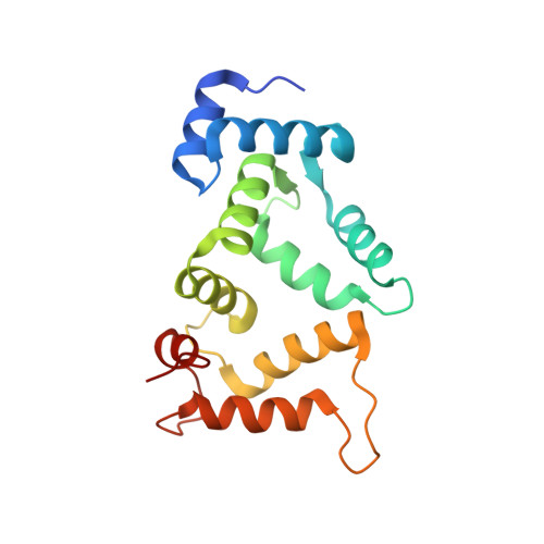
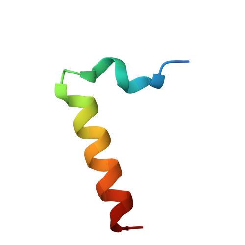
The neuronal calcium sensor NCS-1 regulates the phosphorylation state and activity of the G alpha chaperone and GEF Ric-8A.

Munoz-Reyes, D.McClelland, L.J.Arroyo-Urea, S.Sanchez-Yepes, S.Sabin, J.Perez-Suarez, S.Menendez, M.Mansilla, A.Garcia-Nafria, J.Sprang, S.Sanchez-Barrena, M.J.

(2023) Elife 12

  • DOI: https://doi.org/10.7554/eLife.86151
  • Primary Citation Related Structures: 
    8AHY, 8ALH, 8ALM

  • PubMed Abstract: 

    The neuronal calcium sensor 1 (NCS-1), an EF-hand Ca 2+ binding protein, and Ric-8A coregulate synapse number and probability of neurotransmitter release. Recently, the structures of Ric-8A bound to Gα have revealed how Ric-8A phosphorylation promotes Gα recognition and activity as a chaperone and guanine nucleotide exchange factor. However, the molecular mechanism by which NCS-1 regulates Ric-8A activity and its interaction with Gα subunits is not well understood. Given the interest in the NCS-1/Ric-8A complex as a therapeutic target in nervous system disorders, it is necessary to shed light on this molecular mechanism of action at atomic level. We have reconstituted NCS-1/Ric-8A complexes to conduct a multimodal approach and determine the sequence of Ca 2+ signals and phosphorylation events that promote the interaction of Ric-8A with Gα. Our data show that the binding of NCS-1 and Gα to Ric-8A are mutually exclusive. Importantly, NCS-1 induces a structural rearrangement in Ric-8A that traps the protein in a conformational state that is inaccessible to casein kinase II-mediated phosphorylation, demonstrating one aspect of its negative regulation of Ric-8A-mediated G-protein signaling. Functional experiments indicate a loss of Ric-8A guanine nucleotide exchange factor (GEF) activity toward Gα when complexed with NCS-1, and restoration of nucleotide exchange activity upon increasing Ca 2+ concentration. Finally, the high-resolution crystallographic data reported here define the NCS-1/Ric-8A interface and will allow the development of therapeutic synapse function regulators with improved activity and selectivity.


  • Organizational Affiliation
    • Department of Crystallography and Structural Biology, Institute of Physical-Chemistry 'Blas Cabrera', CSIC, Madrid, Spain.

Macromolecule Content 

  • Total Structure Weight: 24.6 kDa 
  • Atom Count: 1,812 
  • Modeled Residue Count: 199 
  • Deposited Residue Count: 207 
  • Unique protein chains: 2

Macromolecules

Find similar proteins by:|  3D Structure
Entity ID: 1
MoleculeChains  Sequence LengthOrganismDetailsImage
Neuronal calcium sensor 1A [auth B]177Homo sapiensMutation(s): 0 
Gene Names: NCS1FLUPFREQ
UniProt & NIH Common Fund Data Resources
Find proteins for P62166 (Homo sapiens)
Explore P62166 
Go to UniProtKB:  P62166
PHAROS:  P62166
GTEx:  ENSG00000107130 
Entity Groups
Sequence Clusters30% Identity50% Identity70% Identity90% Identity95% Identity100% Identity
UniProt GroupP62166
Sequence Annotations
Expand
Reference Sequence
Find similar proteins by:|  3D Structure
Entity ID: 2
MoleculeChains  Sequence LengthOrganismDetailsImage
Resistance to inhibitors of cholinesterase 8 homolog A (C. elegans)B [auth P]30Rattus norvegicusMutation(s): 0 
UniProt
Find proteins for Q80ZG1 (Rattus norvegicus)
Explore Q80ZG1 
Go to UniProtKB:  Q80ZG1
Entity Groups
Sequence Clusters30% Identity50% Identity70% Identity90% Identity95% Identity100% Identity
UniProt GroupQ80ZG1
Sequence Annotations
Expand
Reference Sequence

Small Molecules

Ligands 6 Unique
IDChains Name / Formula / InChI Key2D Diagram3D Interactions
1PE

Query on 1PE


Download:Ideal Coordinates CCD File
G [auth B],
J [auth P],
K [auth P]
PENTAETHYLENE GLYCOL
C10 H22 O6
JLFNLZLINWHATN-UHFFFAOYSA-N
GOL

Query on GOL


Download:Ideal Coordinates CCD File
H [auth B]GLYCEROL
C3 H8 O3
PEDCQBHIVMGVHV-UHFFFAOYSA-N
CA

Query on CA


Download:Ideal Coordinates CCD File
C [auth B],
D [auth B]
CALCIUM ION
Ca
BHPQYMZQTOCNFJ-UHFFFAOYSA-N
CL

Query on CL


Download:Ideal Coordinates CCD File
I [auth P]CHLORIDE ION
Cl
VEXZGXHMUGYJMC-UHFFFAOYSA-M
MG

Query on MG


Download:Ideal Coordinates CCD File
F [auth B]MAGNESIUM ION
Mg
JLVVSXFLKOJNIY-UHFFFAOYSA-N
NA

Query on NA


Download:Ideal Coordinates CCD File
E [auth B]SODIUM ION
Na
FKNQFGJONOIPTF-UHFFFAOYSA-N

Experimental Data & Validation

Experimental Data

  • Method: X-RAY DIFFRACTION
  • Resolution: 1.86 Å
  • R-Value Free:  0.231 (Depositor), 0.233 (DCC) 
  • R-Value Work:  0.196 (Depositor), 0.196 (DCC) 
  • R-Value Observed: 0.198 (Depositor) 
Space Group: P 41 21 2
Unit Cell:
Length ( Å )Angle ( ˚ )
a = 56.861α = 90
b = 56.861β = 90
c = 134.612γ = 90
Software Package:
Software NamePurpose
PHENIXrefinement
PDB_EXTRACTdata extraction
AutoProcessdata reduction
PHASERphasing
Aimlessdata scaling

Structure Validation

View Full Validation Report


Entry History 

& Funding Information

Deposition Data


Funding OrganizationLocationGrant Number
Ministerio de Ciencia e Innovacion (MCIN)SpainPID2019-111737RB-I00

Revision History  (Full details and data files)

  • Version 1.0: 2023-12-13
    Type: Initial release
  • Version 1.1: 2024-01-10
    Changes: Database references
  • Version 1.2: 2026-03-04
    Changes: Refinement description, Structure summary