PAC-FragmentDEL - photoactivated covalent capture of DNA-encoded fragments for hit discovery.
Ma, H., Murray, J.B., Luo, H., Cheng, X., Chen, Q., Song, C., Duan, C., Tan, P., Zhang, L., Liu, J., Morgan, B.A., Li, J., Wan, J., Baker, L.M., Finnie, W., Guetzoyan, L., Harris, R., Hendrickson, N., Matassova, N., Simmonite, H., Smith, J., Hubbard, R.E., Liu, G.(2022) RSC Med Chem 13: 1341-1349
- PubMed: 36426238
- DOI: https://doi.org/10.1039/d2md00197g
- Primary Citation of Related Structures:
8AHE, 8AHF, 8AHG, 8AHH, 8AHI - PubMed Abstract:
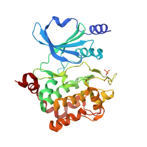
We describe a novel approach for screening fragments against a protein that combines the sensitivity of DNA-encoded library technology with the ability of fragments to explore what will bind. Each of the members of the library consists of a fragment which is linked to a photoactivatable diazirine moiety. Split and pool synthesis combines each fragment with a set of linkers with the version of the library reported here containing some 70k different compounds, each with an individual DNA code. Incubation of the library with a protein sample is followed by photoactivation, washing and subsequent PCR and sequencing which allows the individual fragment hits to be identified. We illustrate how the approach allows successful hit fragment identification using only microgram quantities of material for two targets. PAK4 is a kinase for which conventional fragment screening has generated many advance leads. The as yet undrugged target, 2-epimerase, presents a more challenging active site for identification of hit compounds. In both cases, PAC-FragmentDEL identified fragments validated as hits by ligand-observed NMR measurements and crystal structure determination of off-DNA sample binding to the proteins.
- HitGen Inc. Building 6, No. 8 Huigu First East Road, Tianfu International Bio-Town, Shuangliu District Chengdu 610000 Sichuan P. R. China huiyong.ma@hitgen.com hd.luo@hitgen.com xm.cheng@hitgen.com qx.chen@hitgen.com chao.song@hitgen.com cong.duan@hitgen.com ping.tan@hitgen.com lf.zhang@hitgen.com jian.liu@hitgen.com barry.morgan@hitgen.com jin.li@hitgen.com jq.wan@hitgen.com gs.liu@hitgen.com.
Organizational Affiliation:


