


Complexes of epoxide hydrolase from metagenomic source ch65
Isupov, M.N., De Rose, S.A., Mitchell, D., Littlechild, J.A., Parker, E., Ferrandi, E., Guazzelli, E., Monti, D.To be published.
Experimental Data Snapshot
Starting Model: experimental
View more details
Entity ID: 1 | |||||
|---|---|---|---|---|---|
| Molecule | Chains | Sequence Length | Organism | Details | Image |
| Alpha/beta epoxide hydrolase | A [auth AAA], B [auth BBB] | 299 | metagenome | Mutation(s): 0 | 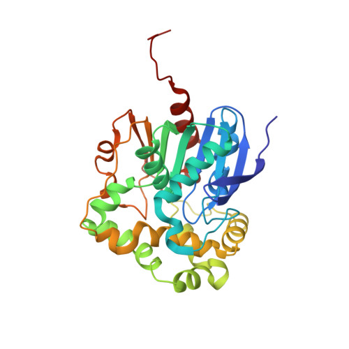 |
| Ligands 5 Unique | |||||
|---|---|---|---|---|---|
| ID | Chains | Name / Formula / InChI Key | 2D Diagram | 3D Interactions | |
| P6G Download:Ideal Coordinates CCD File | M [auth BBB] | HEXAETHYLENE GLYCOL C12 H26 O7 IIRDTKBZINWQAW-UHFFFAOYSA-N |  | ||
| PGE Download:Ideal Coordinates CCD File | E [auth AAA] | TRIETHYLENE GLYCOL C6 H14 O4 ZIBGPFATKBEMQZ-UHFFFAOYSA-N |  | ||
| 2CH (Subject of Investigation/LOI) Download:Ideal Coordinates CCD File | C [auth AAA], N [auth BBB] | 2-CHLOROPHENOL C6 H5 Cl O ISPYQTSUDJAMAB-UHFFFAOYSA-N |  | ||
| EDO Download:Ideal Coordinates CCD File | D [auth AAA] F [auth AAA] G [auth AAA] H [auth AAA] I [auth AAA] | 1,2-ETHANEDIOL C2 H6 O2 LYCAIKOWRPUZTN-UHFFFAOYSA-N |  | ||
| CL Download:Ideal Coordinates CCD File | K [auth AAA], L [auth AAA] | CHLORIDE ION Cl VEXZGXHMUGYJMC-UHFFFAOYSA-M |  | ||
| Length ( Å ) | Angle ( ˚ ) |
|---|---|
| a = 69.21 | α = 90 |
| b = 46.19 | β = 98 |
| c = 92.14 | γ = 90 |
| Software Name | Purpose |
|---|---|
| REFMAC | refinement |
| XDS | data reduction |
| XSCALE | data scaling |
| MOLREP | phasing |
| Coot | model building |
| BUSTER | refinement |
| PARROT | phasing |
| Funding Organization | Location | Grant Number |
|---|---|---|
| European Union (EU) | European Union | 265933 |


