8AG3

Vaccinia C16 N-terminal domains


Experimental Data Snapshot

  • Method: ELECTRON MICROSCOPY
  • Resolution: 3.47 Å
  • Aggregation State: PARTICLE 
  • Reconstruction Method: SINGLE PARTICLE 

wwPDB Validation   3D Report Full Report


This is version 1.1 of the entry. See complete history


Literature

Structural basis for the inactivation of cytosolic DNA sensing by the vaccinia virus.

Rivera-Calzada, A.Arribas-Bosacoma, R.Ruiz-Ramos, A.Escudero-Bravo, P.Boskovic, J.Fernandez-Leiro, R.Oliver, A.W.Pearl, L.H.Llorca, O.

(2022) Nat Commun 13: 7062-7062

  • DOI: https://doi.org/10.1038/s41467-022-34843-z
  • Primary Citation of Related Structures:  
    8AG3, 8AG4, 8AG5

  • PubMed Abstract: 

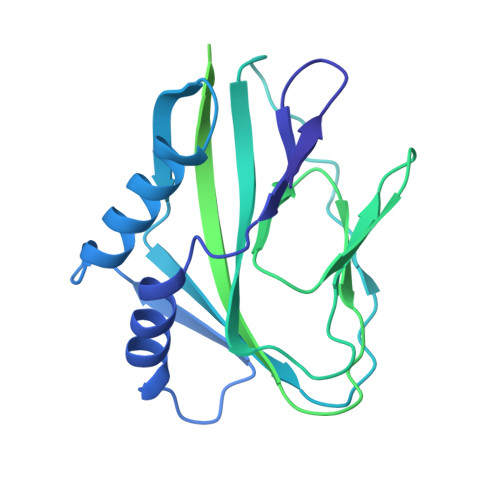
    Detection of cytosolic DNA is a central element of the innate immunity system against viral infection. The Ku heterodimer, a component of the NHEJ pathway of DNA repair in the nucleus, functions as DNA sensor that detects dsDNA of viruses that replicate in the cytoplasm. Vaccinia virus expresses two proteins, C4 and C16, that inactivate DNA sensing and enhance virulence. The structural basis for this is unknown. Here we determine the structure of the C16 - Ku complex using cryoEM. Ku binds dsDNA by a preformed ring but C16 sterically blocks this access route, abrogating binding to a dsDNA end and its insertion into DNA-PK, thereby averting signalling into the downstream innate immunity system. C4 replicates these activities using a domain with 54% identity to C16. Our results reveal how vaccinia virus subverts the capacity of Ku to recognize viral DNA.


  • Organizational Affiliation

    Spanish National Cancer Research Centre (CNIO), Melchor Fernández Almagro 3, 28029, Madrid, Spain.


Macromolecules
Find similar proteins by:  (by identity cutoff)  |  3D Structure
Entity ID: 1
MoleculeChains Sequence LengthOrganismDetailsImage
Protein C10A [auth C],
B [auth D]
369Vaccinia virus Western ReserveMutation(s): 0 
Gene Names: VACWR010C10LVACWR209
UniProt
Find proteins for P03296 (Vaccinia virus (strain Western Reserve))
Explore P03296 
Go to UniProtKB:  P03296
Entity Groups  
Sequence Clusters30% Identity50% Identity70% Identity90% Identity95% Identity100% Identity
UniProt GroupP03296
Sequence Annotations
Expand
  • Reference Sequence
Experimental Data & Validation

Experimental Data

  • Method: ELECTRON MICROSCOPY
  • Resolution: 3.47 Å
  • Aggregation State: PARTICLE 
  • Reconstruction Method: SINGLE PARTICLE 
EM Software:
TaskSoftware PackageVersion
RECONSTRUCTIONRELION
MODEL REFINEMENTPHENIX

Structure Validation

View Full Validation Report


Entry History & Funding Information

Deposition Data


Funding OrganizationLocationGrant Number
Agencia Estatal de Investigacion (AEI)SpainAEI/10.13039/501100011033
Ministerio de Ciencia e Innovacion (MCIN)SpainPID2020-114429RB-I00
Autonomous Community of MadridSpainY2018/BIO-4747
Autonomous Community of MadridSpainP2018/NMT-4443
Cancer Research UKUnited KingdomC302/A14532
Cancer Research UKUnited KingdomC302/A24386

Revision History  (Full details and data files)

  • Version 1.0: 2022-11-09
    Type: Initial release
  • Version 1.1: 2022-11-30
    Changes: Database references