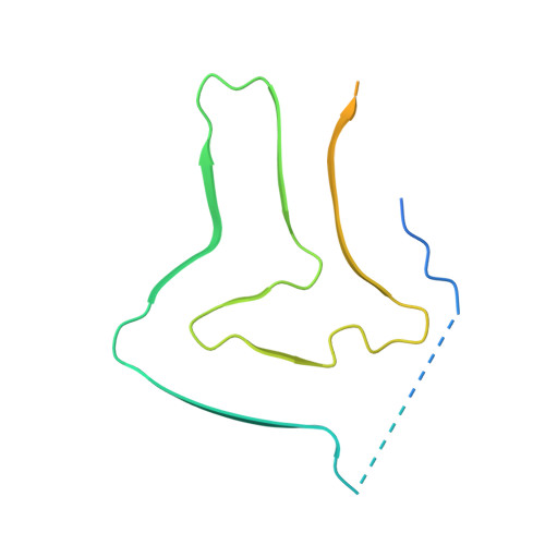
The 3D structure of lipidic fibrils of alpha-synuclein.
Frieg, B., Antonschmidt, L., Dienemann, C., Geraets, J.A., Najbauer, E.E., Matthes, D., de Groot, B.L., Andreas, L.B., Becker, S., Griesinger, C., Schroder, G.F.(2022) Nat Commun 13: 6810-6810
- PubMed: 36357403 Search on PubMedSearch on PubMed Central
- DOI: https://doi.org/10.1038/s41467-022-34552-7
- Primary Citation Related Structures:
8A4L, 8ADS, 8ADU, 8ADV, 8ADW, 8AEX - PubMed Abstract:
α-synuclein misfolding and aggregation into fibrils is a common feature of α-synucleinopathies, such as Parkinson's disease, in which α-synuclein fibrils are a characteristic hallmark of neuronal inclusions called Lewy bodies. Studies on the composition of Lewy bodies extracted postmortem from brain tissue of Parkinson's patients revealed that lipids and membranous organelles are also a significant component. Interactions between α-synuclein and lipids have been previously identified as relevant for Parkinson's disease pathology, however molecular insights into their interactions have remained elusive. Here we present cryo-electron microscopy structures of six α-synuclein fibrils in complex with lipids, revealing specific lipid-fibril interactions. We observe that phospholipids promote an alternative protofilament fold, mediate an unusual arrangement of protofilaments, and fill the central cavities of the fibrils. Together with our previous studies, these structures also indicate a mechanism for fibril-induced lipid extraction, which is likely to be involved in the development of α-synucleinopathies. Specifically, one potential mechanism for the cellular toxicity is the disruption of intracellular vesicles mediated by fibrils and oligomers, and therefore the modulation of these interactions may provide a promising strategy for future therapeutic interventions.
- Institute of Biological Information Processing (IBI-7: Structural Biochemistry) and JuStruct: Jülich Center for Structural Biology, Forschungszentrum Jülich, Jülich, Germany.
Organizational Affiliation: