8AED | pdb_00008aed

Broadly neutralizing DARPin bnD.9 in complex with the HIV-1 envelope variable loop 3 peptide V3 (BG505)


Experimental Data Snapshot

  • Method: X-RAY DIFFRACTION
  • Resolution: 1.17 Å
  • R-Value Free: 
    0.184 (Depositor), 0.184 (DCC) 
  • R-Value Work: 
    0.154 (Depositor), 0.155 (DCC) 
  • R-Value Observed: 
    0.156 (Depositor) 

Starting Model: experimental
View more details

wwPDB Validation 3D Report Full Report

Validation slider image for 8AED

This is version 1.4 of the entry. See complete history

Literature

Trapping the HIV-1 V3 loop in a helical conformation enables broad neutralization.

Glogl, M.Friedrich, N.Cerutti, G.Lemmin, T.Kwon, Y.D.Gorman, J.Maliqi, L.Mittl, P.R.E.Hesselman, M.C.Schmidt, D.Weber, J.Foulkes, C.Dingens, A.S.Bylund, T.Olia, A.S.Verardi, R.Reinberg, T.Baumann, N.S.Rusert, P.Dreier, B.Shapiro, L.Kwong, P.D.Pluckthun, A.Trkola, A.

(2023) Nat Struct Mol Biol 30: 1323-1336

  • DOI: https://doi.org/10.1038/s41594-023-01062-z
  • Primary Citation Related Structures: 
    7TXD, 7Z7C, 8AED

  • PubMed Abstract: 

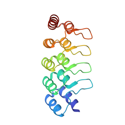
    The third variable (V3) loop on the human immunodeficiency virus 1 (HIV-1) envelope glycoprotein trimer is indispensable for virus cell entry. Conformational masking of V3 within the trimer allows efficient neutralization via V3 only by rare, broadly neutralizing glycan-dependent antibodies targeting the closed prefusion trimer but not by abundant antibodies that access the V3 crown on open trimers after CD4 attachment. Here, we report on a distinct category of V3-specific inhibitors based on designed ankyrin repeat protein (DARPin) technology that reinstitute the CD4-bound state as a key neutralization target with up to >90% breadth. Broadly neutralizing DARPins (bnDs) bound V3 solely on open envelope and recognized a four-turn amphipathic α-helix in the carboxy-terminal half of V3 (amino acids 314-324), which we termed 'αV3C'. The bnD contact surface on αV3C was as conserved as the CD4 binding site. Molecular dynamics and escape mutation analyses underscored the functional relevance of αV3C, highlighting the potential of αV3C-based inhibitors and, more generally, of postattachment inhibition of HIV-1.


  • Organizational Affiliation
    • Institute for Medical Virology, University of Zurich (UZH), Zurich, Switzerland.

Macromolecule Content 

  • Total Structure Weight: 40.38 kDa 
  • Atom Count: 3,411 
  • Modeled Residue Count: 362 
  • Deposited Residue Count: 372 
  • Unique protein chains: 2

Macromolecules

Find similar proteins by:|  3D Structure
Entity ID: 1
MoleculeChains  Sequence LengthOrganismDetailsImage
Broadly neutralizing DARPin bnD.9
A, B
162synthetic constructMutation(s): 0 
Find similar proteins by:  Sequence   |   3D Structure  
Entity ID: 2
MoleculeChains  Sequence LengthOrganismDetailsImage
Envelope glycoprotein gp160
C, D
24Human immunodeficiency virus 1Mutation(s): 0 
UniProt
Find proteins for Q9Q714 (Human immunodeficiency virus type 1 group M subtype H (isolate VI991))
Explore Q9Q714 
Go to UniProtKB:  Q9Q714
Entity Groups
Sequence Clusters30% Identity50% Identity70% Identity90% Identity95% Identity100% Identity
UniProt GroupQ9Q714
Sequence Annotations
Expand
Reference Sequence

Small Molecules

Ligands 4 Unique
IDChains Name / Formula / InChI Key2D Diagram3D Interactions
CIT

Query on CIT


Download:Ideal Coordinates CCD File
J [auth A],
N [auth B]
CITRIC ACID
C6 H8 O7
KRKNYBCHXYNGOX-UHFFFAOYSA-N
EDO

Query on EDO


Download:Ideal Coordinates CCD File
E [auth A],
F [auth A],
K [auth B],
L [auth B]
1,2-ETHANEDIOL
C2 H6 O2
LYCAIKOWRPUZTN-UHFFFAOYSA-N
ACT

Query on ACT


Download:Ideal Coordinates CCD File
G [auth A],
H [auth A]
ACETATE ION
C2 H3 O2
QTBSBXVTEAMEQO-UHFFFAOYSA-M
MG

Query on MG


Download:Ideal Coordinates CCD File
I [auth A],
M [auth B],
O [auth C]
MAGNESIUM ION
Mg
JLVVSXFLKOJNIY-UHFFFAOYSA-N

Experimental Data & Validation

Experimental Data

  • Method: X-RAY DIFFRACTION
  • Resolution: 1.17 Å
  • R-Value Free:  0.184 (Depositor), 0.184 (DCC) 
  • R-Value Work:  0.154 (Depositor), 0.155 (DCC) 
  • R-Value Observed: 0.156 (Depositor) 
Space Group: P 1 21 1
Unit Cell:
Length ( Å )Angle ( ˚ )
a = 50.795α = 90
b = 55.233β = 98.54
c = 58.032γ = 90
Software Package:
Software NamePurpose
PHENIXrefinement
PDB_EXTRACTdata extraction
autoPROCdata reduction
STARANISOdata scaling
PHASERphasing

Structure Validation

View Full Validation Report


Entry History 

& Funding Information

Deposition Data


Funding OrganizationLocationGrant Number
Swiss National Science FoundationSwitzerland--

Revision History  (Full details and data files)

  • Version 1.0: 2023-08-16
    Type: Initial release
  • Version 1.1: 2023-08-23
    Changes: Database references
  • Version 1.2: 2023-08-30
    Changes: Database references
  • Version 1.3: 2023-09-20
    Changes: Database references
  • Version 1.4: 2026-03-04
    Changes: Refinement description, Structure summary