8ADF

X-ray crystal structure of PPAR gamma ligand binding domain in complex with CZ39


Experimental Data Snapshot

  • Method: X-RAY DIFFRACTION
  • Resolution: 2.13 Å
  • R-Value Free: 0.257 
  • R-Value Work: 0.216 
  • R-Value Observed: 0.219 

Starting Model: experimental
View more details

wwPDB Validation   3D Report Full Report


Ligand Structure Quality Assessment 


This is version 1.2 of the entry. See complete history


Literature

Biological Screening and Crystallographic Studies of Hydroxy gamma-Lactone Derivatives to Investigate PPAR gamma Phosphorylation Inhibition.

Capelli, D.Cazzaniga, G.Mori, M.Laghezza, A.Loiodice, F.Quaglia, M.Negro, E.Meneghetti, F.Villa, S.Montanari, R.

(2023) Biomolecules 13

  • DOI: https://doi.org/10.3390/biom13040694
  • Primary Citation of Related Structures:  
    8ADF, 8C0C

  • PubMed Abstract: 

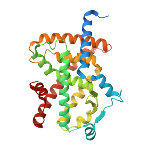
    PPARγ represents a key target for the treatment of type 2 diabetes and metabolic syndrome. To avoid serious adverse effects related to the PPARγ agonism profile of traditional antidiabetic drugs, a new opportunity is represented by the development of molecules acting as inhibitors of PPARγ phosphorylation by the cyclin-dependent kinase 5 (CDK5). Their mechanism of action is mediated by the stabilization of the PPARγ β-sheet containing Ser273 (Ser245 in PPARγ isoform 1 nomenclature). In this paper, we report the identification of new γ-hydroxy-lactone-based PPARγ binders from the screening of an in-house library. These compounds exhibit a non-agonist profile towards PPARγ, and one of them prevents Ser245 PPARγ phosphorylation by acting mainly on PPARγ stabilization and exerting a weak CDK5 inhibitory effect.


  • Organizational Affiliation

    Istituto di Cristallografia, Consiglio Nazionale delle Ricerche, Strada Provinciale 35d, n. 9-00010, Montelibretti, 34149 Rome, Italy.


Macromolecules
Find similar proteins by:  (by identity cutoff)  |  3D Structure
Entity ID: 1
MoleculeChains Sequence LengthOrganismDetailsImage
Peroxisome proliferator-activated receptor gamma
A, B
303Homo sapiensMutation(s): 0 
Gene Names: PPARGNR1C3
UniProt & NIH Common Fund Data Resources
Find proteins for P37231 (Homo sapiens)
Explore P37231 
Go to UniProtKB:  P37231
PHAROS:  P37231
GTEx:  ENSG00000132170 
Entity Groups  
Sequence Clusters30% Identity50% Identity70% Identity90% Identity95% Identity100% Identity
UniProt GroupP37231
Sequence Annotations
Expand
  • Reference Sequence
Small Molecules
Ligands 1 Unique
IDChains Name / Formula / InChI Key2D Diagram3D Interactions
LRA
Query on LRA

Download Ideal Coordinates CCD File 
C [auth A],
D [auth B]
(2R)-3-(4-bromophenyl)-2-(3-hydroxyphenyl)-4-oxidanyl-2H-furan-5-one
C16 H11 Br O4
AXUOPMIBBDYMEV-OAHLLOKOSA-N
Experimental Data & Validation

Experimental Data

  • Method: X-RAY DIFFRACTION
  • Resolution: 2.13 Å
  • R-Value Free: 0.257 
  • R-Value Work: 0.216 
  • R-Value Observed: 0.219 
  • Space Group: C 1 2 1
Unit Cell:
Length ( Å )Angle ( ˚ )
a = 92.878α = 90
b = 59.954β = 103.29
c = 117.37γ = 90
Software Package:
Software NamePurpose
PHENIXrefinement
PDB_EXTRACTdata extraction
XDSdata reduction
XSCALEdata scaling
PHASERphasing

Structure Validation

View Full Validation Report


Ligand Structure Quality Assessment 


Entry History & Funding Information

Deposition Data


Funding OrganizationLocationGrant Number
Not funded--

Revision History  (Full details and data files)

  • Version 1.0: 2023-04-26
    Type: Initial release
  • Version 1.1: 2023-05-24
    Changes: Database references
  • Version 1.2: 2024-02-07
    Changes: Data collection, Refinement description