8ACS

Crystal structure of FMO from Janthinobacterium svalbardensis


Experimental Data Snapshot

  • Method: X-RAY DIFFRACTION
  • Resolution: 2.50 Å
  • R-Value Free: 0.229 
  • R-Value Work: 0.182 
  • R-Value Observed: 0.184 

wwPDB Validation   3D Report Full Report


Ligand Structure Quality Assessment 


This is version 1.3 of the entry. See complete history


Literature

A Cold-Active Flavin-Dependent Monooxygenase from Janthinobacterium svalbardensis Unlocks Applications of Baeyer-Villiger Monooxygenases at Low Temperature.

Chanique, A.M.Polidori, N.Sovic, L.Kracher, D.Assil-Companioni, L.Galuska, P.Parra, L.P.Gruber, K.Kourist, R.

(2023) ACS Catal 13: 3549-3562

  • DOI: https://doi.org/10.1021/acscatal.2c05160
  • Primary Citation of Related Structures:  
    8ACS

  • PubMed Abstract: 

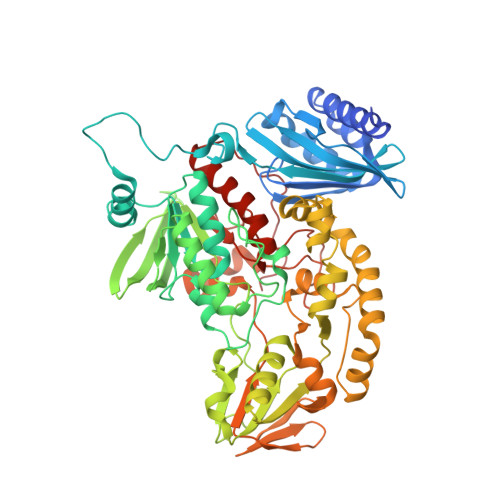
    Cold-active enzymes maintain a large part of their optimal activity at low temperatures. Therefore, they can be used to avoid side reactions and preserve heat-sensitive compounds. Baeyer-Villiger monooxygenases (BVMO) utilize molecular oxygen as a co-substrate to catalyze reactions widely employed for steroid, agrochemical, antibiotic, and pheromone production. Oxygen has been described as the rate-limiting factor for some BVMO applications, thereby hindering their efficient utilization. Considering that oxygen solubility in water increases by 40% when the temperature is decreased from 30 to 10 °C, we set out to identify and characterize a cold-active BVMO. Using genome mining in the Antarctic organism Janthinobacterium svalbardensis, a cold-active type II flavin-dependent monooxygenase (FMO) was discovered. The enzyme shows promiscuity toward NADH and NADPH and high activity between 5 and 25 °C. The enzyme catalyzes the monooxygenation and sulfoxidation of a wide range of ketones and thioesters. The high enantioselectivity in the oxidation of norcamphor (eeS = 56%, eeP > 99%, E > 200) demonstrates that the generally higher flexibility observed in the active sites of cold-active enzymes, which compensates for the lower motion at cold temperatures, does not necessarily reduce the selectivity of these enzymes. To gain a better understanding of the unique mechanistic features of type II FMOs, we determined the structure of the dimeric enzyme at 2.5 Å resolution. While the unusual N-terminal domain has been related to the catalytic properties of type II FMOs, the structure shows a SnoaL-like N-terminal domain that is not interacting directly with the active site. The active site of the enzyme is accessible only through a tunnel, with Tyr-458, Asp-217, and His-216 as catalytic residues, a combination not observed before in FMOs and BVMOs.


  • Organizational Affiliation

    NAWI Graz, BioTechMed-Graz, Institute of Molecular Biotechnology, Graz University of Technology, Petersgasse 14, Graz 8010, Austria.


Macromolecules
Find similar proteins by:  (by identity cutoff)  |  3D Structure
Entity ID: 1
MoleculeChains Sequence LengthOrganismDetailsImage
FAD-dependent oxidoreductase
A, B, C, D
618Janthinobacterium svalbardensisMutation(s): 0 
Gene Names: CNX70_19775
UniProt
Find proteins for A0A290WZ30 (Janthinobacterium svalbardensis)
Explore A0A290WZ30 
Go to UniProtKB:  A0A290WZ30
Entity Groups  
Sequence Clusters30% Identity50% Identity70% Identity90% Identity95% Identity100% Identity
UniProt GroupA0A290WZ30
Sequence Annotations
Expand
  • Reference Sequence
Small Molecules
Ligands 3 Unique
IDChains Name / Formula / InChI Key2D Diagram3D Interactions
FAD (Subject of Investigation/LOI)
Query on FAD

Download Ideal Coordinates CCD File 
E [auth A],
J [auth B],
P [auth C],
V [auth D]
FLAVIN-ADENINE DINUCLEOTIDE
C27 H33 N9 O15 P2
VWWQXMAJTJZDQX-UYBVJOGSSA-N
PEG
Query on PEG

Download Ideal Coordinates CCD File 
I [auth A],
K [auth B],
R [auth C],
W [auth D],
X [auth D]
DI(HYDROXYETHYL)ETHER
C4 H10 O3
MTHSVFCYNBDYFN-UHFFFAOYSA-N
GOL
Query on GOL

Download Ideal Coordinates CCD File 
F [auth A]
G [auth A]
H [auth A]
L [auth B]
M [auth B]
F [auth A],
G [auth A],
H [auth A],
L [auth B],
M [auth B],
N [auth B],
O [auth B],
Q [auth C],
S [auth C],
T [auth C],
U [auth C]
GLYCEROL
C3 H8 O3
PEDCQBHIVMGVHV-UHFFFAOYSA-N
Experimental Data & Validation

Experimental Data

  • Method: X-RAY DIFFRACTION
  • Resolution: 2.50 Å
  • R-Value Free: 0.229 
  • R-Value Work: 0.182 
  • R-Value Observed: 0.184 
  • Space Group: P 21 21 2
Unit Cell:
Length ( Å )Angle ( ˚ )
a = 142.302α = 90
b = 316.361β = 90
c = 65.647γ = 90
Software Package:
Software NamePurpose
autoPROCdata processing
XDSdata reduction
PHENIXrefinement
Aimlessdata scaling
PHASERphasing

Structure Validation

View Full Validation Report


Ligand Structure Quality Assessment 


Entry History & Funding Information

Deposition Data


Funding OrganizationLocationGrant Number
Austrian Science FundAustriaDOC-46

Revision History  (Full details and data files)

  • Version 1.0: 2022-09-07
    Type: Initial release
  • Version 1.1: 2023-03-08
    Changes: Database references
  • Version 1.2: 2023-04-05
    Changes: Database references
  • Version 1.3: 2024-05-01
    Changes: Data collection, Refinement description